Li Huang1, Radhouene Neji1,2, Filippo Bosio1, Amedeo Chiribiri1, Reza Razavi1, and Sébastien Roujol1
1School of Biomedical Engineering and Imaging Sciences, Faculty of Life Sciences and Medicine, King's College London, London, United Kingdom, 2MR Research Collaborations, Siemens Healthcare Limited, Frimley, United Kingdom
1School of Biomedical Engineering and Imaging Sciences, Faculty of Life Sciences and Medicine, King's College London, London, United Kingdom, 2MR Research Collaborations, Siemens Healthcare Limited, Frimley, United Kingdom
ECV mapping with
time-efficient full left ventricular coverage can be achieved using FAST1 at
1.5T yielding limited penalty of ECV spatial variability and highly correlated
ECV values in comparison with MOLLI.
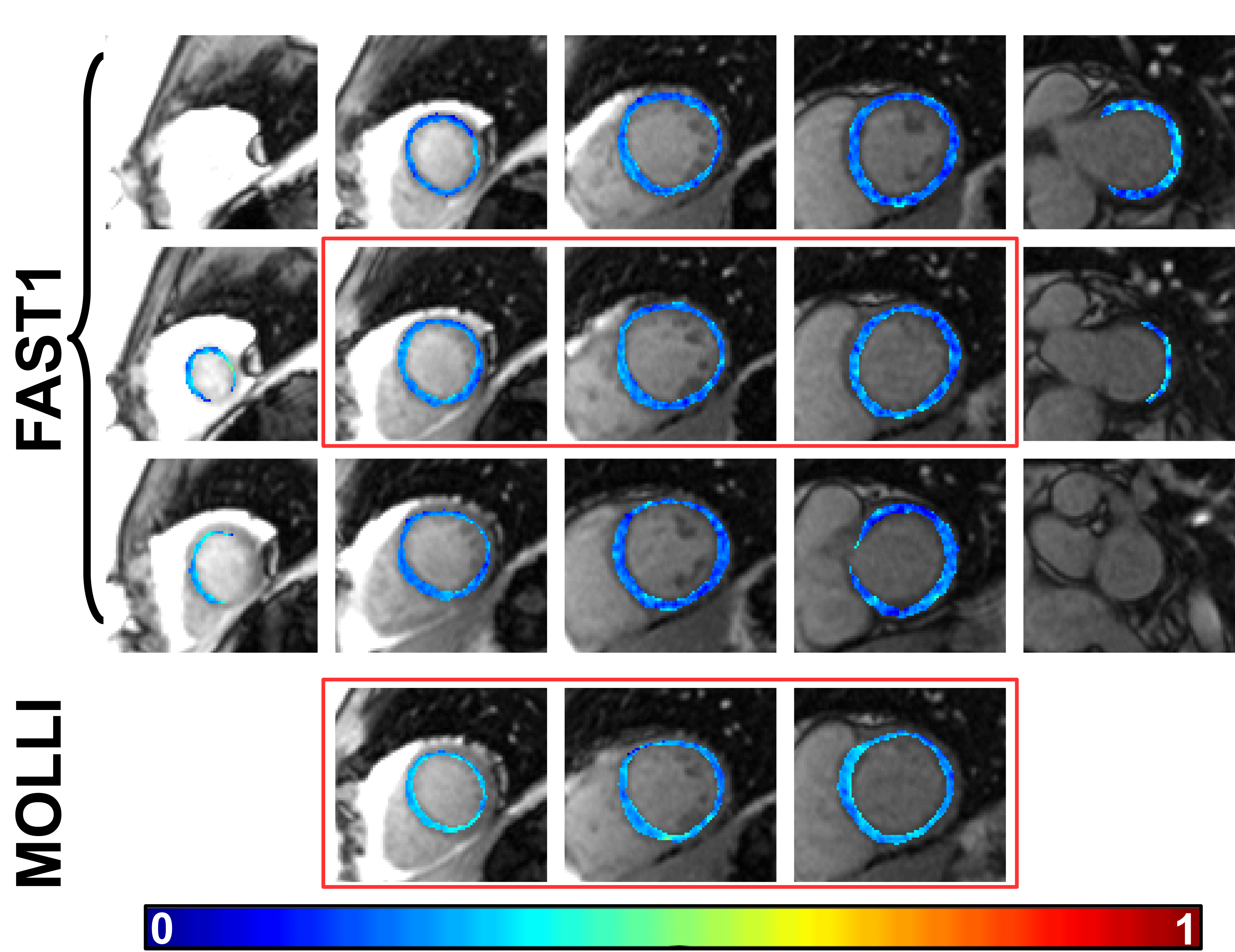
Fig. 1. Representative example ECV maps of a
patient admitted for suspected myocarditis using FAST1 and MOLLI. Note that each row
of FAST1 T1 maps and each MOLLI T1 map represent a sequence run within one
breathhold, respectively. The red rectangles indicate the three common slice positions in
both datasets.
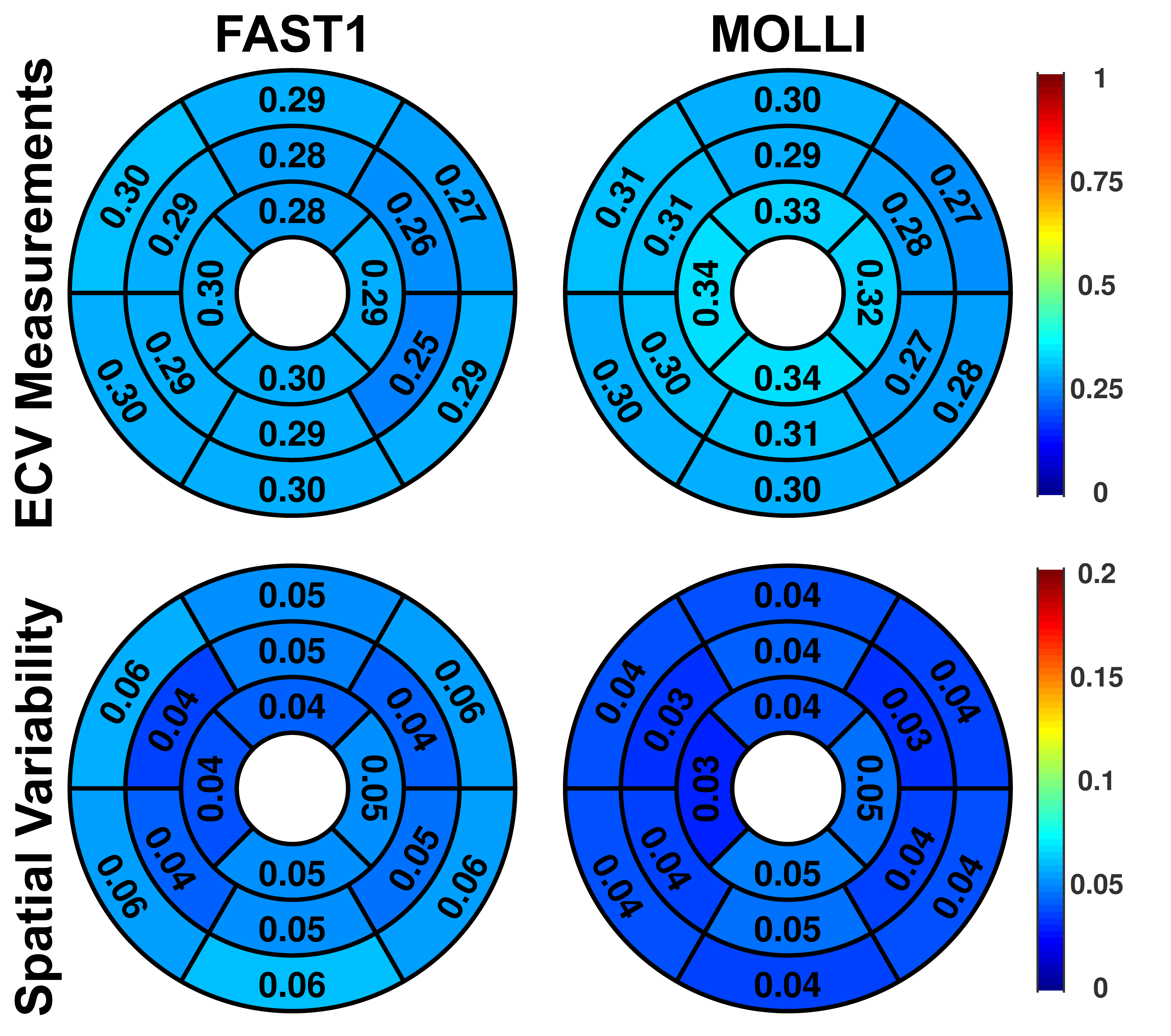
Fig. 3. Segmental analysis of FAST1 and MOLLI
regarding to ECV measurements and spatial variability across all patients.