Ashitha Pathrose1, Roberto Sarnari1, Sabeth Essl1, Carson Herman1, Daniel Gordon1, Kelvin Chow1,2, Benjamin Freed3, Michael Cuttica3, Michael Markl1,4, and James Carr1,3,4
1Radiology, Northwestern University, Chicago, IL, United States, 2Siemens Medical Solutions USA, Chicago, IL, United States, 3Medicine, Northwestern University, Chicago, IL, United States, 4Biomedical Engineering, Northwestern University, Chicago, IL, United States
1Radiology, Northwestern University, Chicago, IL, United States, 2Siemens Medical Solutions USA, Chicago, IL, United States, 3Medicine, Northwestern University, Chicago, IL, United States, 4Biomedical Engineering, Northwestern University, Chicago, IL, United States
CMR-derived left ventricular feature-tracking strain, native
T1, and tissue velocities were significantly different in pulmonary
hypertensive patients when compared to controls. These measures also correlated
significantly with right heart catheterization derived pressures.
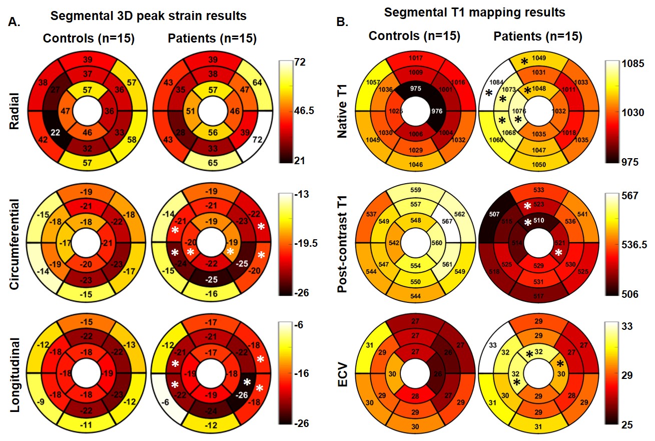
Figure
1. Schematic representation of the strain and T1 mapping results. (A.) shows increased
circumferential and longitudinal peak strains at the mid-septal region and LV
free wall; (B.) shows increased native T1 times mainly on the septal and
anterior LV myocardium and few segmental differences in the post-contrast T1
times and ECV (based on 16-segment AHA model; * indicates significant
difference, p<0.05)
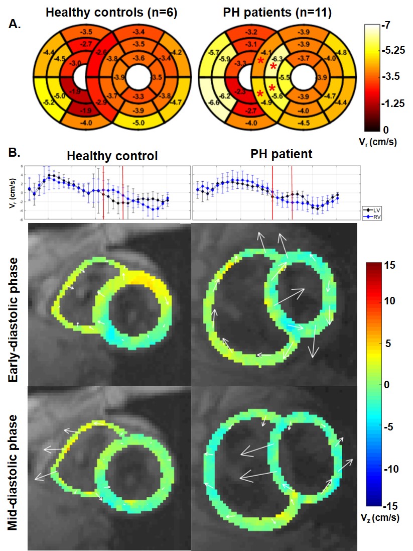
Figure
2. Schematic representation of the TPM results. (A.) segmental radial peak
diastolic velocities averaged for the controls and patients (based on extended 16+10
segment AHA model; * indicates significant difference, p<0.05); (B.) early
and mid-diastolic in-plane velocity (represented as vectors) for a representative
control and PH patient