Verónica Aramendía-Vidaurreta1, Pedro Macías-Gordaliza2,3, Marta Vidorreta4, Rebeca Echeverria-Chasco1, Gorka Bastarrika1, Arrate Muñoz-Barrutia2,3, and María Fernández-Seara1
1Radiology, Clínica Universidad de Navarra, Pamplona, Spain, 2Universidad Carlos III de Madrid, Madrid, Spain, 3Instituto de Investigación Sanitaria Gregorio Marañón, Madrid, Spain, 4Siemens Healthineers, Madrid, Spain
1Radiology, Clínica Universidad de Navarra, Pamplona, Spain, 2Universidad Carlos III de Madrid, Madrid, Spain, 3Instituto de Investigación Sanitaria Gregorio Marañón, Madrid, Spain, 4Siemens Healthineers, Madrid, Spain
In
this work, we demonstrate the feasibility of quantifying MBF employing synchronized
breathing techniques with FAIR-ASL in healthy subjects. Motion is significantly
reduced, MBF values are consistent with literature and coefficient of variation
for intrasession reproducibility is 13%.

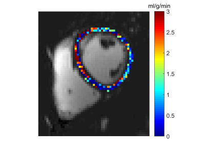
Figure 2: MBF map of a representative
volunteer for a FAIR-ASL sequence and synchronized breathing acquisitions overlapped to the mean ASL image. Average
MBF within myocardium is 1.12 ml/g/min.

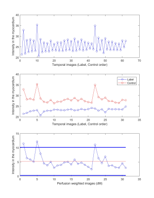
Figure 1. Top row
shows mean signal intensity within myocardium for all the temporal image series, where label and
control acquisitions are alternated. Middle row shows signal intensity within the myocardium
for label (blue) and control (red) series independently. Bottom row depicts
the mean ASL signal in the myocardium, showing high tSNR (3.45). Red line indicates the mean across all pairs. Blue lines indicate
two standard deviations from the ASL mean within the myocardium before
excluding outliers. Then, three images were identified as outliers and discarded.
