Jeesoo Lee1,2, Nadia El Hangouche2, Alex J Barker3, James D Thomas2, and Michael Markl1,4
1Radiology, Northwestern University, Chicago, IL, United States, 2Cardiology, Northwestern Memorial Hospital, Chicago, IL, United States, 3Radiology, University of Colorado Denver, Denver, CO, United States, 4Biomedical Engineering, Northwestern University, Chicago, IL, United States
1Radiology, Northwestern University, Chicago, IL, United States, 2Cardiology, Northwestern Memorial Hospital, Chicago, IL, United States, 3Radiology, University of Colorado Denver, Denver, CO, United States, 4Biomedical Engineering, Northwestern University, Chicago, IL, United States
A novel framework was developed that integrates 4D flow MRI and color Doppler echocardiography for direct assessment of intra-cardiac velocity measured by both modalities. This framework allowed the investigation of inter-modal agreement in the anatomical region-of-interest.
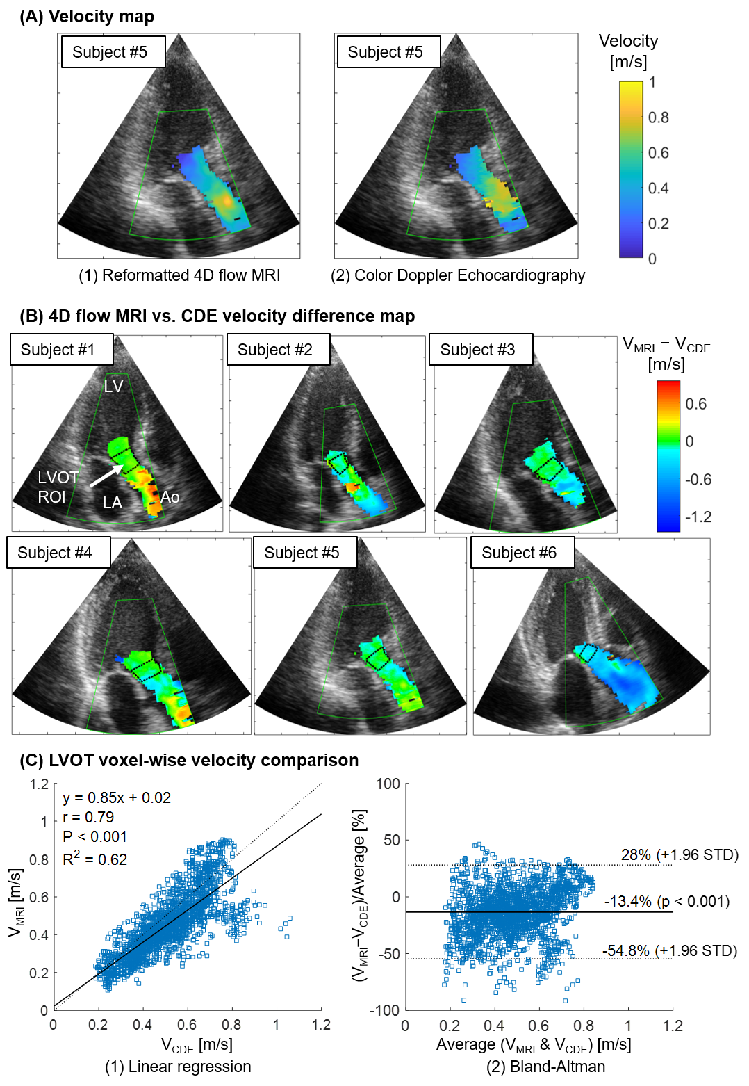
Figure 4. Voxel-wise velocity comparison results.
(A1) Reformatted 4D flow MRI velocity, VMRI, map and (A2) original
color Doppler echocardiography (CDE) velocity, VCDE map from the subject
#5. (B) Peak systole velocity difference (VMRI−VCDE) map from
all subjects. Black dotted line box represents the left ventricular outflow
tract (LVOT) region. (C1) Linear regression plot with the identity
curve (dotted line) and (C2) Bland-Altman analysis results for LVOT velocity
comparison. All subjects are included in the analysis.
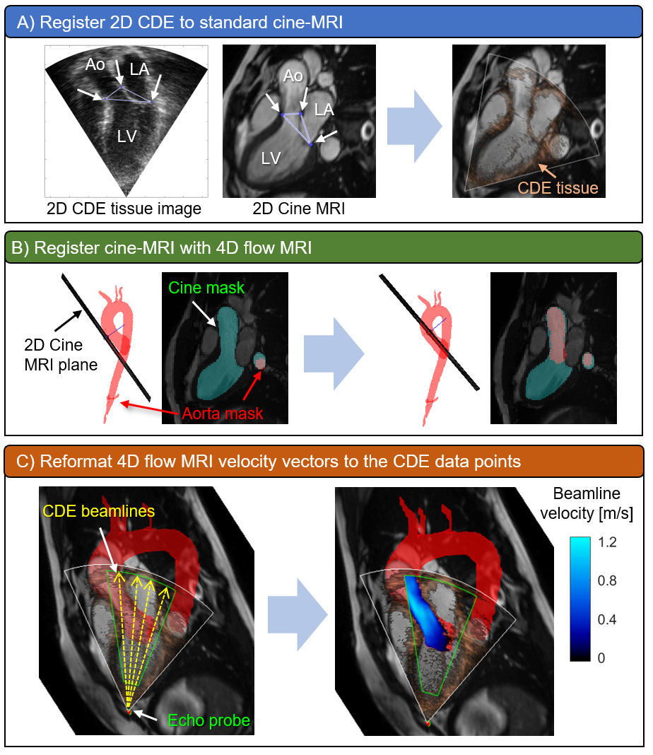
Figure 1. 4D flow MRI and color Doppler
echocardiography (CDE) data integration workflow. (A) A CDE tissue image was
manually registered to a 3C cine MRI by matching anatomical landmarks (white arrows).
(B) The cine image was registered with 4D flow MRI by
matching the segmentation masks. (C) 4D flow MRI velocity vectors were
interpolated to CDE pixels and projected to ultrasound beamlines for
comparison.