Judith Zimmermann1,2,3, Kathrin Bäumler1, Michael Loecher1,3, Alison Marsden4,5, and Daniel Ennis1,3,5
1Radiology, Stanford University, Stanford, CA, United States, 2Computer Science, Technical University of Munich, Munich, Germany, 3Radiology, Veterans Administration Health Care System, Palo Alto, CA, United States, 4Pediatrics, Stanford University, Stanford, CA, United States, 5Cardiovascular Institute, Stanford, CA, United States
1Radiology, Stanford University, Stanford, CA, United States, 2Computer Science, Technical University of Munich, Munich, Germany, 3Radiology, Veterans Administration Health Care System, Palo Alto, CA, United States, 4Pediatrics, Stanford University, Stanford, CA, United States, 5Cardiovascular Institute, Stanford, CA, United States
Feasibility
of flow measurements in a 3D-printed subject-specific thoracic aorta is shown. Choosing
appropriate vessel wall thickness and compliance for models is essential in
quantitative 4D-flow MRI studies.
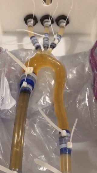
Fig. 2. Animation
of model M1_1.6_soft embedded into the flow circuit with HR = 60/min and a
total flow volume of 4.38 l/min. The brown fluid color is caused by the use of
ferumoxytol contrast agent (0.75ml per liter of fluid). Pulsatile motion of the
wall can be observed.
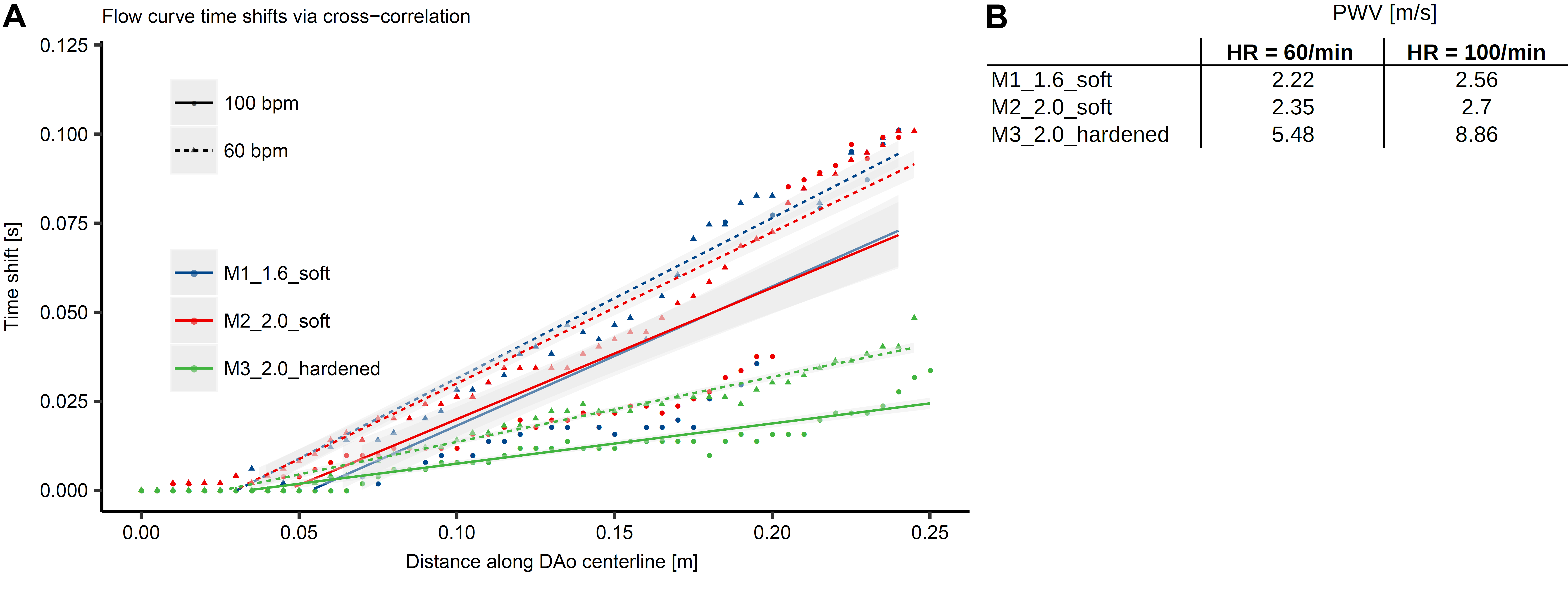
Fig. 5. PWV
estimation along the descending aorta centerline. A: Measured time-shift over distance points and linear regression of
the time-shift over distance along the centerline for three models at two heart
rates (triangles/dashed lines for HR=60, dots/solid lines for HR=100). B: Derived PWV values, i.e. inverse
slope values for each fitted linear regression line, increase with increasing stiffness,
increasing wall thickness, and higher heart rate.