Anqin Li1, Zhen Li1, and Daoyu Hu1
1Tongji Hospital, Tongji Medical College, Huazhong University of Science and Technology, Wuhan, China
1Tongji Hospital, Tongji Medical College, Huazhong University of Science and Technology, Wuhan, China
MRI could be useful in improving the assessment
of complex renal cysts.
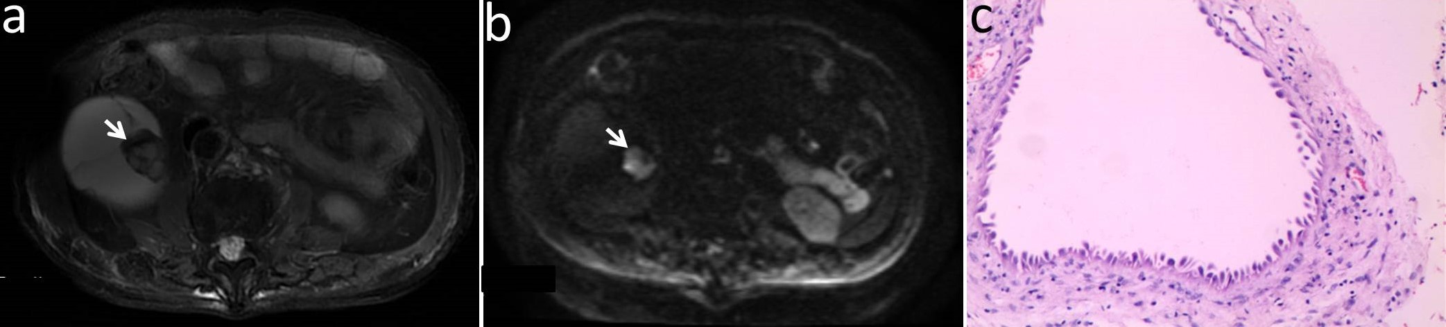
Figure 4: Images in a 78-year-old man with a cystic mass in the right kidney. (a) Axial T2-weighted MR
image shown mural nodular soft tissue (arrow); (b) Axial DWI (b = 1000 s/mm2) image shown remarkably
high signal intensity of the wall nodule (arrow); (c) This lesion was
surgically removed and determined to be a multilocular cystic renal neoplasm of
low malignant potential.
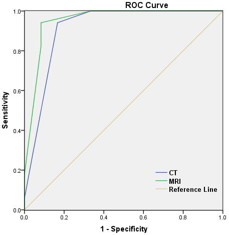
Figure 1: Receiver
operating characteristic (ROC) curves of the characteristics of lesions on CT
and MRI images for differentiating benign renal cystic masses from malignant.
ROC curves of MRI in differentiating benign from malignant lesions generated
a higher AUC (AUC, 0.951; Sensitivity, 94.1%; Specificity, 91.7%) than CT (AUC, 0.912; Sensitivity, 94.1%;
Specificity, 83.3%).