Xianyun Cai1, Guangbin Wang2, and Jinxia Zhu3
1radiology, Shandong Medical Imaging Research Institute, Shandong University, jinan, China, 2Shandong Medical Imaging Research Institute, Shandong University, jinan, China, 3MR Collaboration, Siemens Healthcare Ltd., beijing, China
1radiology, Shandong Medical Imaging Research Institute, Shandong University, jinan, China, 2Shandong Medical Imaging Research Institute, Shandong University, jinan, China, 3MR Collaboration, Siemens Healthcare Ltd., beijing, China
This study explored the scanning strategy of fetal
spine imaging using Magnetic Resonance Imaging (MRI) on 315 volunteer pregnant
subjects. Whole-spine MRI was performed on fetuses using Susceptibility-Weighted
Imaging (SWI) and True Fast Imaging with Steady-state Precession (TrueFISP)
sequences. Images from both methods were acquired, and the diagnostic
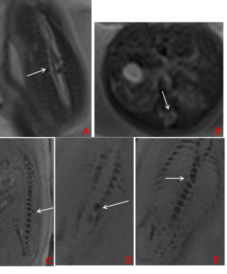
efficacy was compared. The SWI showed superior performance in visualizing osseous spinal anomalies, while
TrueFISP better presented spinal canal contents lesions. Additionally,
the MRI was preferred for the diagnosis of fetal spinal diseases to US. These
mixed results suggest that a combination of both techniques is appropriate for
fetal spine imaging


