Akshay Chaudhari1, Quin Lu2, Anna Wisser3,4, Wolfgang Wirth3,4, Garry E Gold1, Brian A Hargreaves1, and Felix Eckstein3,4
1Stanford University, Stanford, CA, United States, 2Philips, San Francisco, CA, United States, 3Paracelsus Medical University, Salzburg, Austria, 4Chondrometrics GmbH, Ainring, Germany
1Stanford University, Stanford, CA, United States, 2Philips, San Francisco, CA, United States, 3Paracelsus Medical University, Salzburg, Austria, 4Chondrometrics GmbH, Ainring, Germany
Here we show qDESS harmonization
across vendors, which can produce high scan-rescan repeatability and high
repeatability of left-right knee asymmetry for cartilage volume, surface area,
and thickness metrics.

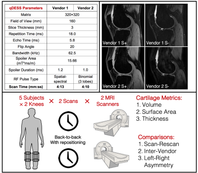
Figure
1: An overall
schematic of the study depicting the scan parameters of the qDESS sequence
across the two vendors (top left) and the two echo contrasts (S+ and S-) that
qDESS generates, that are favorable for cartilage segmentation (top right).
(Bottom) The study design shows that both knees from 5 subjects were scanned separately
twice each, on 2 different scanners. Cartilage volume, surface area, and thickness
was computed and these metrics were used to compare scan-rescan and inter-vendor variations along with left-right knee asymmetry.

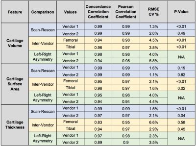
Figure
2:
A quantitative
summary of all comparisons performed in evaluating the scan-rescan variation
(blue), inter-vendor variation (yellow), and left-right asymmetry (green) for cartilage volume, surface area, and thickness. Agreement between
all metrics was high, as demonstrated using the concordance and Pearson
correlation coefficients, and RMSE-CV%. For the scan-rescan and inter-vendor
metrics that were significantly different, the RMSE-CV% was low, which suggests only a
small, but systematic bias, likely due to partial volume effects of 3mm slices.