Nadia D. Bragagnolo1,2, Benjamin J. Geraghty1,2, Casey Y. Lee1,2, Albert P. Chen3, and Charles H. Cunningham1,2
1Medical Biophysics, University of Toronto, Toronto, ON, Canada, 2Physical Sciences, Sunnybrook Research Institute, Toronto, ON, Canada, 3GE Healthcare Technologies, Toronto, ON, Canada
1Medical Biophysics, University of Toronto, Toronto, ON, Canada, 2Physical Sciences, Sunnybrook Research Institute, Toronto, ON, Canada, 3GE Healthcare Technologies, Toronto, ON, Canada
B1+ measurements acquired at multiple locations immediately prior to imaging are used to derive power input scaling factors for each slice. These scaling factors ensure that the desired flip angle is achieved across a large field-of-view for multi-organ imaging.

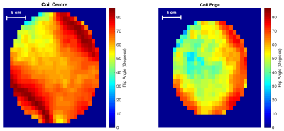
Two slices from a B1+ map of the flexible 13C volume transmit-receive coil surrounding a circular phantom (diameter = 26 cm). These are axial slices obtained using the double angle method with a target flip angle of 60°. Prior to mapping, the power input was calibrated to the left slice, directly through the centre of the coil. This calibration resulted in the induced flip angle of the right slice, 6 cm away near the coil edge, being significantly lower than the desired 60°.

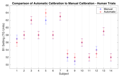
Results comparing the rapid B1+ measurement sequence to manual B1+ measurements during human 13C scans (n = 14). Transmit gain (TG) is the console variable used to scale the power input to the coil, controlling the magnitude of B1+ generated. For a given subject, the rapid measure was within ±1μT of what was found manually.