Jasmine Y. Graham1,2, Peder E.Z. Larson2, Daniel B. Vigneron2, and Jeremy W. Gordon2
1Bioengineering, UC San Francisco, UC Berkeley, San Francisco, CA, United States, 2Radiology and Biomedical Imaging, UC San Francisco, San Francisco, CA, United States
1Bioengineering, UC San Francisco, UC Berkeley, San Francisco, CA, United States, 2Radiology and Biomedical Imaging, UC San Francisco, San Francisco, CA, United States
Utilizing a multi-resolution
simulation framework, lactate
and bicarbonate images have improved SNR of 3.3-fold and 5.7-fold compared to
high resolution images. Kinetic rate fits for multi-resolution images
accurately estimate ground truth values.
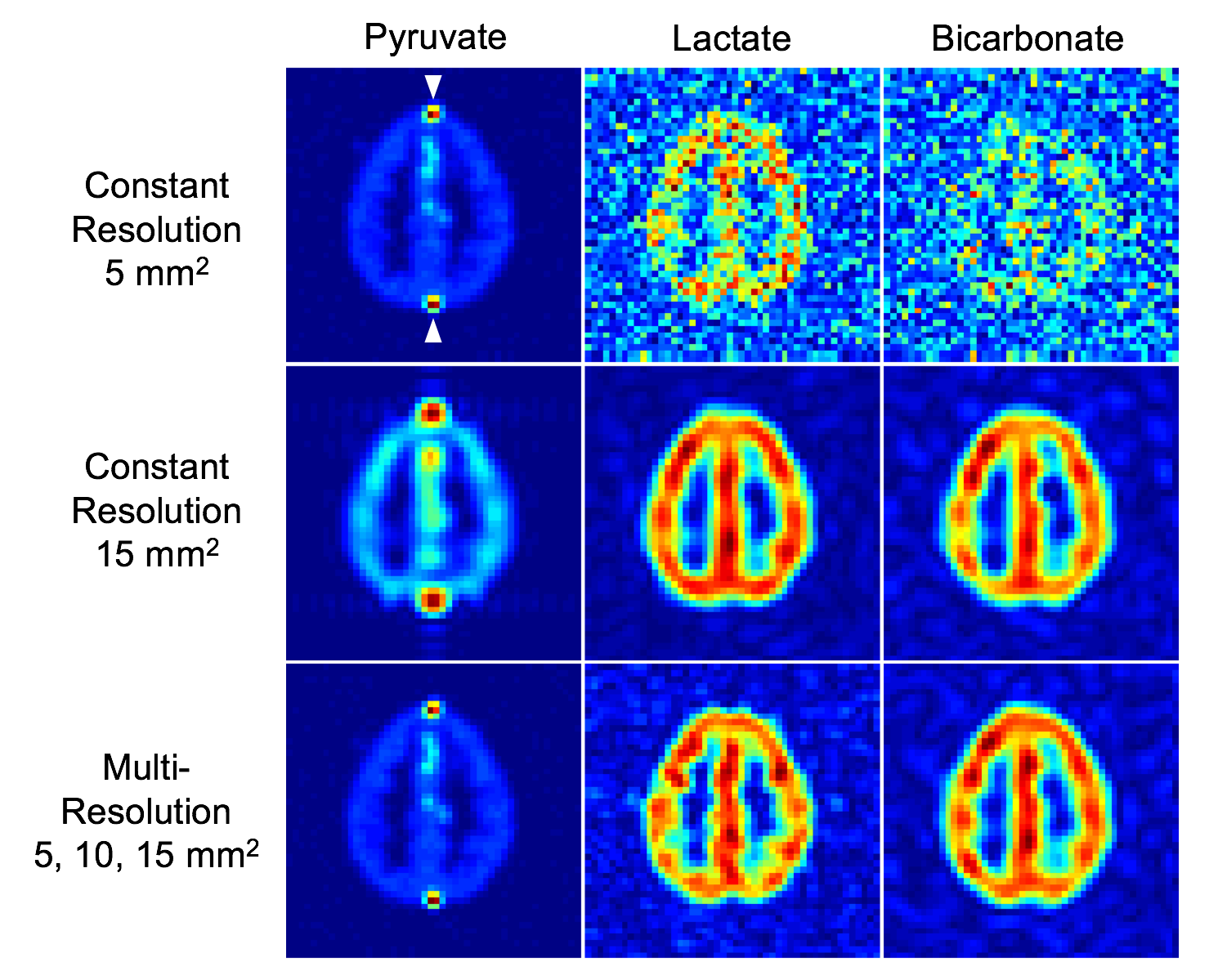
Figure 2. Simulated brain phantoms
of hyperpolarized [1-C13]pyruvate, lactate, and bicarbonate signals
from 30 seconds after pyruvate bolus delivery. The white arrows in the top left
image indicate vascular ROIs with high pyruvate signal and no lactate or
bicarbonate signal. The multi-resolution images of lactate (10ⅹ10 mm2) and bicarbonate (15ⅹ15 mm2) have a respective mean SNR gain of 3.3-fold and 5.7-fold
compared to the constant high resolution (5ⅹ5 mm2) images.
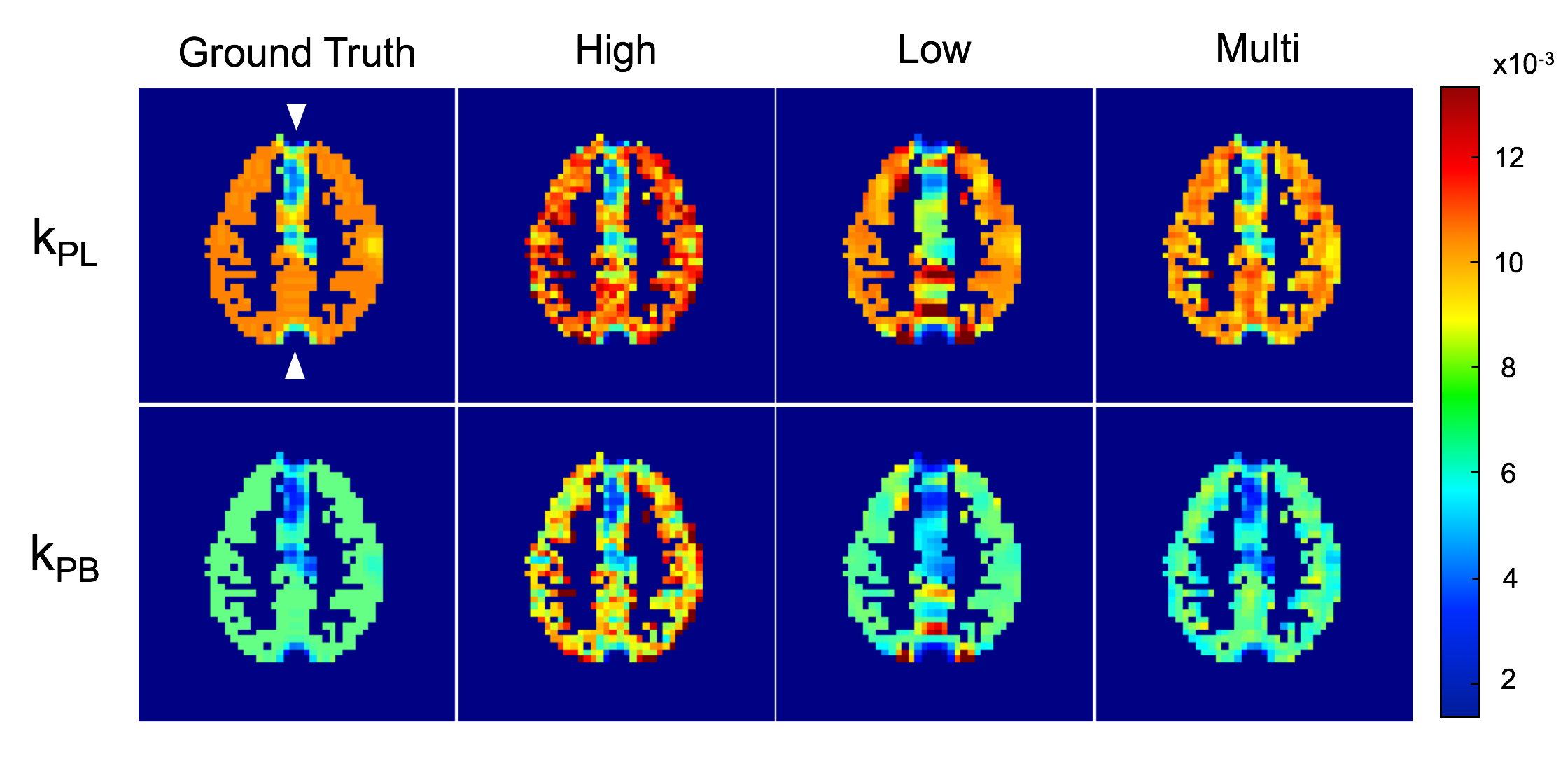
Figure 3. Kinetic rate maps for
pyruvate to lactate and pyruvate to bicarbonate using images with different
resolution schemes. The zero magnitude areas as indicated by the white arrows
in the top left panel are vascular ROIs with high pyruvate signal and no conversion
to lactate or bicarbonate. The multi-resolution maps yield the most accurate estimations
of the true kinetic rates.