Timothy J Allen1, Leah C Henze Bancroft2, Manoj Kumar2, Tyler Bradshaw2, Roberta M Strigel1,2,3, Alan McMillan1,2, and Amy M Fowler1,2,3
1Medical Physics, University of Wisconsin-Madison, Madison, WI, United States, 2Radiology, University of Wisconsin-Madison, Madison, WI, United States, 3Carbone Cancer Center, University of Wisconsin-Madison, Madison, WI, United States
1Medical Physics, University of Wisconsin-Madison, Madison, WI, United States, 2Radiology, University of Wisconsin-Madison, Madison, WI, United States, 3Carbone Cancer Center, University of Wisconsin-Madison, Madison, WI, United States
SUVmean and SUVmax
values in breast cancers, benign fibroglandular tissue, descending
aorta, and liver did not change following injection of a gadolinium-based
contrast agent. These results suggest that GBCAs are unlikely to affect PET
quantitation in simultaneous PET/MR breast imaging.
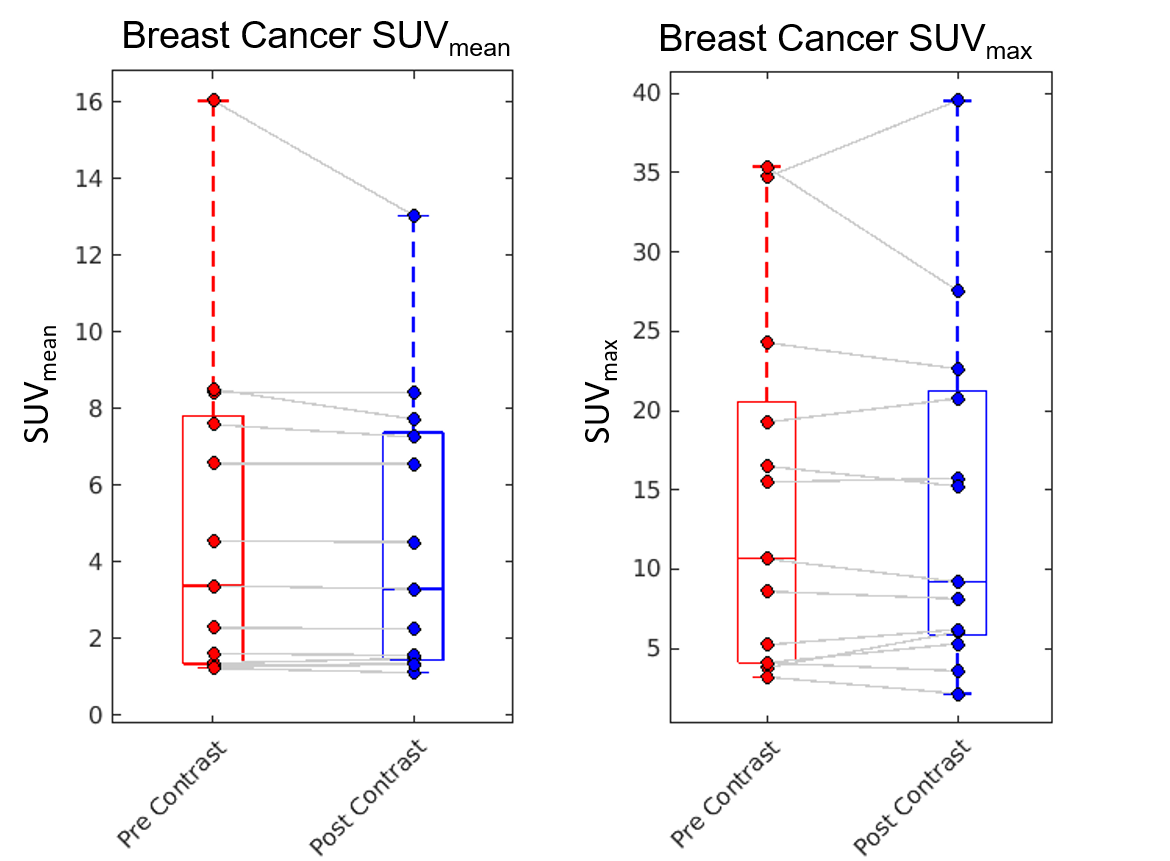
Figure 2: SUVmean and
SUVmax in known breast cancers does not significantly change
following administration of a gadolinium based contrast agent.
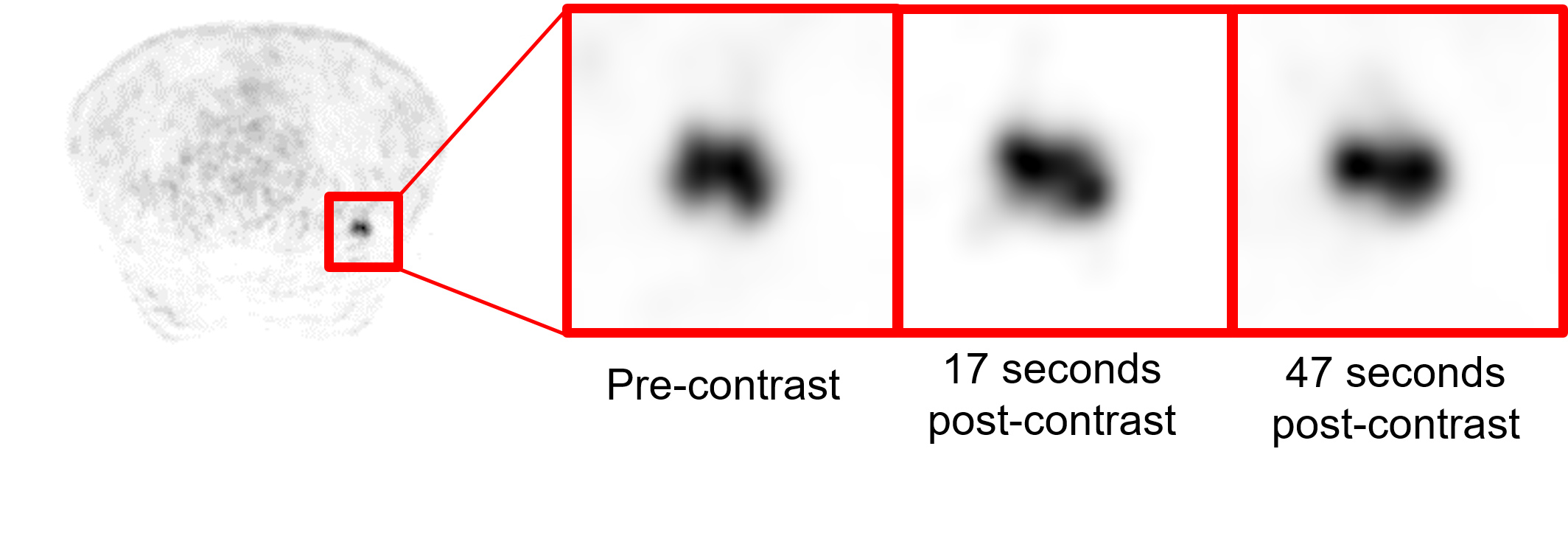
Figure 1: PET images pre- and post-injection of a gadolinium
based contrast agent demonstrating FDG uptake in a biopsy-proven breast cancer
in the posterior left breast. No visual change can be perceived between
pre-contrast images and post-contrast images. 30 second dynamic reconstructions
of PET data are shown for a pre-contrast frame and 2 post-contrast frames.