Steffen Goerke1, Johannes Breitling1, Katharina M. Kubera2, Moritz Zaiss3, Dusan Hirjak4, Robert C. Wolf2, Anoshirwan A. Tavakoli5, Heinz-Peter Schlemmer5, Daniel Paech5, Mark E. Ladd1, and Peter Bachert1
1Divsion of Medical Physics in Radiology, German Cancer Research Center (DKFZ), Heidelberg, Germany, 2Center for Psychosocial Medicine, Department of General Psychiatry, Heidelberg University, Heidelberg, Germany, 3Neuroradiology, University of Erlangen-Nürnberg, Erlangen, Germany, 4Departement of Psychiatry and Psychotherapy, Central Institute of Mental Health, Medical Faculty Mannheim, Heidelberg University, Mannheim, Germany, 5Departement of Radiology, German Cancer Research Center (DKFZ), Heidelberg, Germany
1Divsion of Medical Physics in Radiology, German Cancer Research Center (DKFZ), Heidelberg, Germany, 2Center for Psychosocial Medicine, Department of General Psychiatry, Heidelberg University, Heidelberg, Germany, 3Neuroradiology, University of Erlangen-Nürnberg, Erlangen, Germany, 4Departement of Psychiatry and Psychotherapy, Central Institute of Mental Health, Medical Faculty Mannheim, Heidelberg University, Mannheim, Germany, 5Departement of Radiology, German Cancer Research Center (DKFZ), Heidelberg, Germany
A significant decrease of the rNOE-CEST
signal in Alzheimer’s patients was observed supporting the hypothesis that CEST-MRI
allows the diagnostic imaging of patients with Alzheimer's disease.
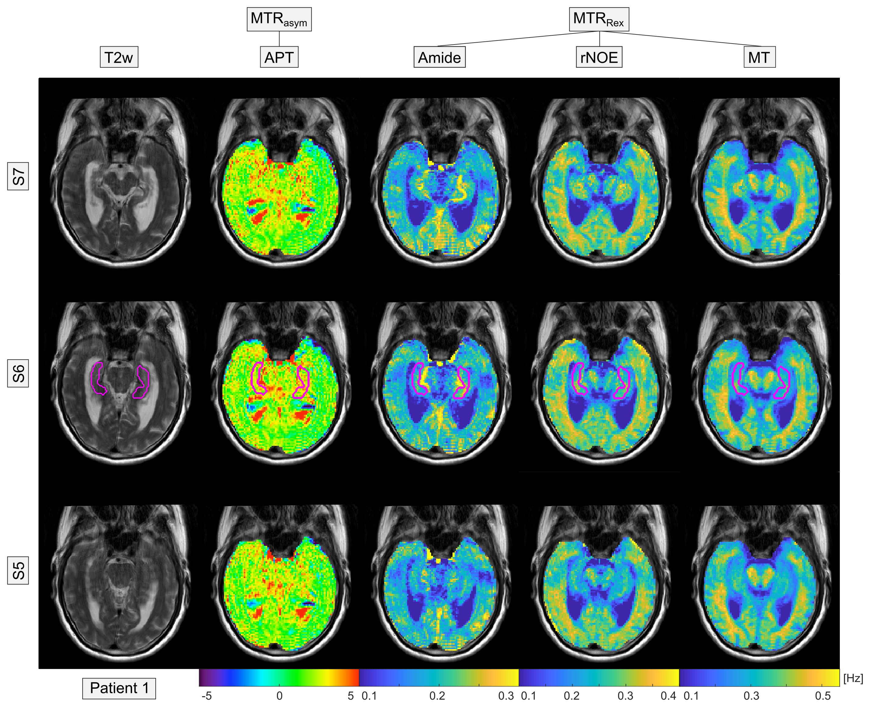
Figure 2: 3D-CEST-MRI (Slice 4-6 of
12) of an exemplary Alzheimer’s patient. Besides conventional APT imaging (MTRasym),
the relaxation-compensated CEST contrasts (MTRRex) of the isolated
Amide, rNOE and MT signal are displayed. For statistical analysis, a ROI in the
central part of the Hippocampus (magenta region) was selected on the T2w image.

Figure 3: Group means of the different
CEST contrasts in the Hippocampus of 4 Alzheimer’s patients and 2 healthy
volunteers. In Alzheimer’s patients a significant decrease, with group mean
values outside the error range, was observed for the rNOE and MT signal. Errors
were calculated using the standard deviation (SD).