Roya Afshari1,2, Francesco Santini1,2, Rahel Heule3, Craig H. Meyer4, Josef Pfeuffer5, and Oliver Bieri1,2
1Division of Radiological Physics,Department of Radiology, University Hospital Basel, University of Basel, Basel, Switzerland, 2Department of Biomedical Engineering, University of Basel, Basel, Switzerland, 3High Field Magnetic Resonance, Max Planck Institute for Biological Cybernetics, Tübingen, Germany, 4Department of Biomedical Engineering, University of Virginia, Charlottesville, VA, United States, 5Siemens Healthcare, Application Development, Erlangen, Germany
1Division of Radiological Physics,Department of Radiology, University Hospital Basel, University of Basel, Basel, Switzerland, 2Department of Biomedical Engineering, University of Basel, Basel, Switzerland, 3High Field Magnetic Resonance, Max Planck Institute for Biological Cybernetics, Tübingen, Germany, 4Department of Biomedical Engineering, University of Virginia, Charlottesville, VA, United States, 5Siemens Healthcare, Application Development, Erlangen, Germany
A one-minute whole-brain
MTR imaging method with intrinsic B1-correction was introduced. The
new method makes use of three interleaved multi-slice acquisitions offering high reproducibility and excellent
prospects for translation and application in the clinics.
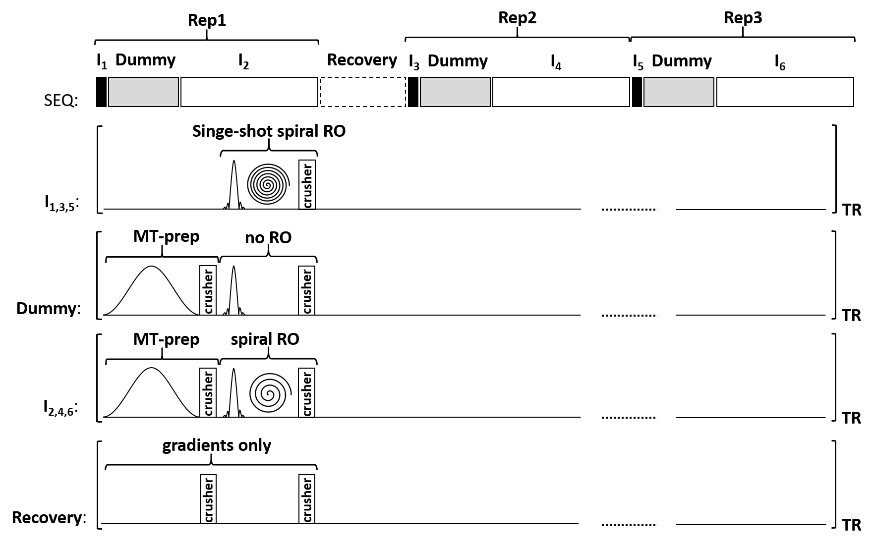
Scheme of 2D
interleaved multi-slice spiral MTR imaging approach. SEQ: the sequence features
three repetitions. Each repetition features a single-shot spiral readout,
followed by a dummy period to reach steady state conditions and an interleaved
spiral readout for image encoding. I1,3,5: Single shot spiral
readout without MT-preparation pulses. Dummy: MT-preparation pulses, RF
excitation and gradients are played out. No readout is performed. I2,4,6:
MT-preparation is played out. Image encoding is performed using spiral
interleaves.
Recovery: Only gradients are played out.
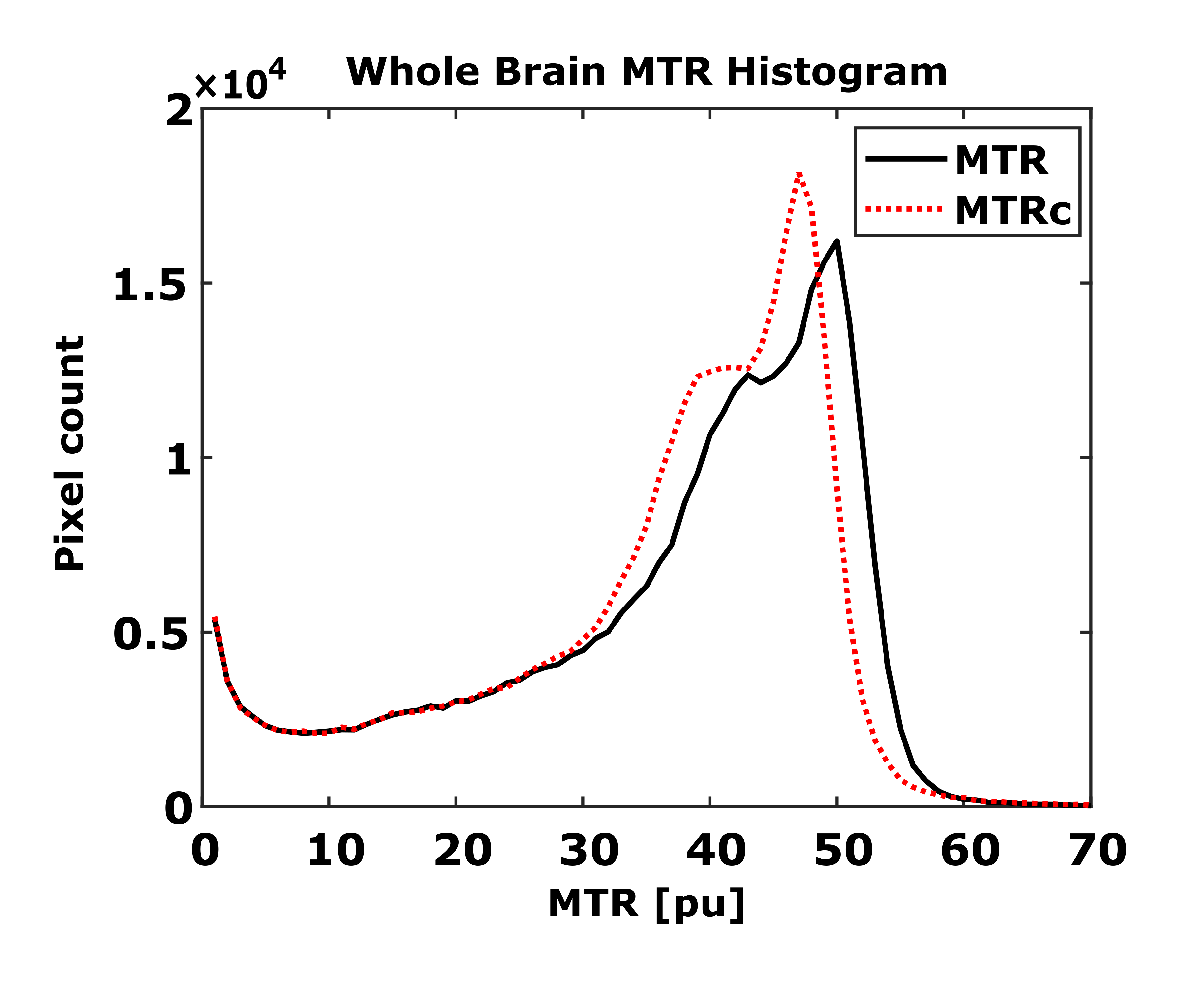
Whole brain MTR
histograms for the B1-uncorrected (black solid line) and for the B1-corrected
MT-saturation (red dotted line). Overall, B1-correction leads to a shift of the
histogram toward lower MTR values, lower spread and improved
peak-discrimination.