Lucas Soustelle1,2, Samira Mchinda1,2, Thomas Troalen3, Maxime Guye1,2, Jean-Philippe Ranjeva1,2, Gopal Varma4, David C Alsop4, Guillaume Duhamel1,2, and Olivier M Girard1,2
1Aix-Marseille Univ, CNRS, CRMBM, Marseille, France, 2APHM, Hôpital Universitaire Timone, CEMEREM, Marseille, France, 3Siemens Healthcare SAS, Saint-Denis, France, 4Division of MR Research, Radiology, Beth Israel Deaconess Medical Center, Harvard Medical School, Boston, MA, United States
1Aix-Marseille Univ, CNRS, CRMBM, Marseille, France, 2APHM, Hôpital Universitaire Timone, CEMEREM, Marseille, France, 3Siemens Healthcare SAS, Saint-Denis, France, 4Division of MR Research, Radiology, Beth Israel Deaconess Medical Center, Harvard Medical School, Boston, MA, United States
As for many other MRI techniques, inhomogeneous Magnetization
Transfer necessitates special attention to local B1+ variations. In this work, a
detailed analysis of the ihMT ratio sensitivity to B1+ is performed on human
brain at 3T using various concentration schemes for its optimization.

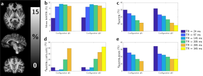
Figure 2: Left side (a):
illustrative 3D ihMTR images (2mm isotropic resolution) obtained under strong
RF energy concentration conditions (TR = 345 ms, Configuration #2). Right side:
WM averaged ihMTR measured at nominal Vref (b), WM averaged metrics calculated
using the two reference voltage settings: δihMTR (ihMTR relative
variation; c), δihMTR-[-10%;10%] (voxel fraction within 10% error
margin; d) and δihMTR-RMS (RMS relative error; e).

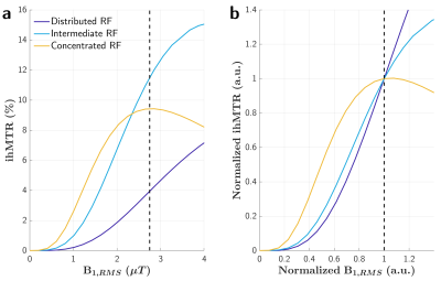
Figure 1: Simulations of the
ihMT ratio in WM as a function of RF power B1,RMS for distributed
(short TR), intermediate and concentrated (long TR) RF power deposition. The
dashed vertical line indicates the nominal B1,RMS used in the
current 3T study. a: absolute ihMTR values show that concentrating RF energy
allows increasing ihMTR sensitivity; b: normalized ihMTR values provide insight
into the corresponding relative sensitivity to B1+ variations (approximated at
first order by the derivative of the ihMTR vs. B1,RMS characteristic
curves at nominal B1,RMS).
