Jinyoung Kim1, Rémi Patriat1, Jordan Kaplan1, and Noam Harel1
1Center for Magnetic Resonance Research, University of Minnesota, Minneapolis, MN, United States
1Center for Magnetic Resonance Research, University of Minnesota, Minneapolis, MN, United States
The
proposed semi-supervised deep context-aware network outperforms the existing
atlas-based segmentation tool and popular deep neural networks in dentate and
interposed nuclei segmentation on 7T diffusion MRIs in terms of accuracy and
consistency.

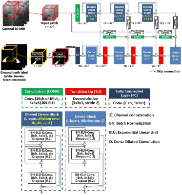
The overall scheme of the proposed
segmentation framework. Sub-volume (patch) based processing reduces the memory burden
and significantly increases the number of training samples. The size of input
and output patches is 32×32×32 and the patch step size is 5×5×5. Dilation rates
D = (1,1,2), (1,1,2,4), and (1,1,2,4,8), respectively, are applied to convolutional
layers of three dilated dense blocks where the number of layers L = 3, 4, and
5. The number of channels in dense blocks increases by 8.

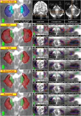
Visual comparison of segmentation
results on a 7T B0 MRI of a specific subject along with measures. The first row
is volumetric ground truth and selected planes. The first column is volumetric
segmentation obtained by each method. The last three columns are corresponding
contours in each plane. Red and green are segmented dentate and interposed
nuclei, blue and light blue are the ground truth dentate and interposed nuclei.
Arrows indicate anterior direction in the axial plane.
