Wei Yu1,2, Lixin Wang1,2, Xianchang Zhang3, Zhentao Zuo4, Rong Xue4,5,6, and Tianyi Qian1,2
1Sinovation Ventures AI Institute, Beijing, China, 2QuantMind, Beijing, China, 3MR Collaboration, Siemens Healthcare, Beijing, China, 4State Key Laboratory of Brain and Cognitive Science, Beijing MRI Center for Brain Research, Institute of Biophysics, Chinese Academy of Sciences, Beijing, China, 5University of Chinese Academy of Sciences, Beijing, China, 6Beijing Institute for Brain Disorders, Beijing, China
1Sinovation Ventures AI Institute, Beijing, China, 2QuantMind, Beijing, China, 3MR Collaboration, Siemens Healthcare, Beijing, China, 4State Key Laboratory of Brain and Cognitive Science, Beijing MRI Center for Brain Research, Institute of Biophysics, Chinese Academy of Sciences, Beijing, China, 5University of Chinese Academy of Sciences, Beijing, China, 6Beijing Institute for Brain Disorders, Beijing, China
Compared with current reconstruction methods, this method
can simultaneously reconstruct well-defined anatomical details and salient
blood vessels. This reconstruction method may be useful for increasing the efficiency
of brain MRI examinations.
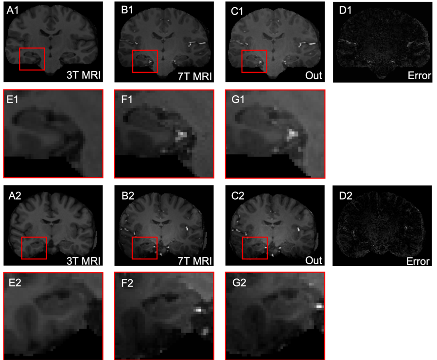
Figure 1: Results of 7T MRI reconstruction using FocalGAN on two testing pairs
shown in coronal view. 3T MRI (A) is the
input, 7T MRI (B) is the reference,
Out (C) designates the reconstruction generated by FocalGAN, error maps (D) are shown in the right-most
column. Regions outlined in red are expanded beneath the relevant panel (E-G).
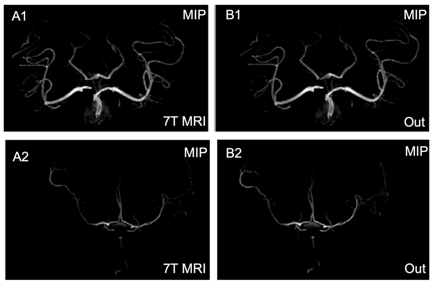
Figure 2: Maximal intensity projection (MIP) images
show improved imaging of blood vessel reconstruction in two test pairs. 7T MRI MIP (A) refers to reference
images, Out MIP (B) designates results
generated using FocalGAN.