Dongxue Li1, Bin Dai1, Zhenliang Xiong1, Xianchun Zeng1, Lisha Nie2, Pu-Yeh Wu2, and Rongpin Wang1
1Department of Radiology, Guizhou Provincial People's Hospital, Guiyang, China, 2GE Healthcare, MR Research, Beijing, China
1Department of Radiology, Guizhou Provincial People's Hospital, Guiyang, China, 2GE Healthcare, MR Research, Beijing, China
Here we introduced a standardized brain region susceptibility quantification procedure based on QSM and Brainnetome Atlas. Our results show that this method has high accuracy in measuring brain iron content.
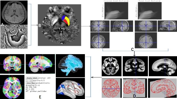
Figure 1. MR-QSM images were registered, smoothed and corresponded to the standard brain by MATLAB software.
B refers to the QSM image obtained by processing the original data(A), and it can be seen that the globus pallidus(red), putamen(yellow) and caudate nucleus(blue) are obtained by manual drawing. Using the MATLAB software, we first registered QSM and susceptibility images of each participant to its own structural image, then normalized to the standard brain, and finally smoothed (C and D). E represents the position of each ROI on the Brainnetome Atlas brain network map.
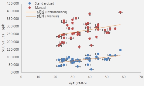
Figure 4. Magnetic susceptibility values (in ppb) measured from standardized and manual ROI selection methods were positively correlated with age.