Nina Pötsch1, Marcus Raudner1,2, Tom Hilbert3,4,5, Tobias Kober3,4,5, Elisabeth Weiland6, Panagiotis Kapetas1, and Pascal Baltzer1
1Department of Biomedical Imaging and Image-guided Therapy, Medical University of Vienna, Vienna, Austria, 2High Field MR Centre, Department of Biomedical Imaging and Image-guided Therapy, Medical University of Vienna, Vienna, Austria, 3Advanced Clinical Imaging Technology, Siemens Healthcare, Lausanne, Switzerland, 4Department of Radiology, Lausanne University Hospital and University of Lausanne, Lausanne, Switzerland, 5LTS5, Ecole Polytechnique Fédérale de Lausanne (EPFL), Lausanne, Switzerland, 6Siemens Healthcare GmbH, Erlangen, Germany
1Department of Biomedical Imaging and Image-guided Therapy, Medical University of Vienna, Vienna, Austria, 2High Field MR Centre, Department of Biomedical Imaging and Image-guided Therapy, Medical University of Vienna, Vienna, Austria, 3Advanced Clinical Imaging Technology, Siemens Healthcare, Lausanne, Switzerland, 4Department of Radiology, Lausanne University Hospital and University of Lausanne, Lausanne, Switzerland, 5LTS5, Ecole Polytechnique Fédérale de Lausanne (EPFL), Lausanne, Switzerland, 6Siemens Healthcare GmbH, Erlangen, Germany
Feasibility study using GRAPPATINI and compressed sensing MP2RAGE
prototype sequences for high-resolution T1/T2 maps of the breast in 7:32 min.
Examples of malignant or benign lesions show initial evidence of the
quantitative value’s clinical relevance.
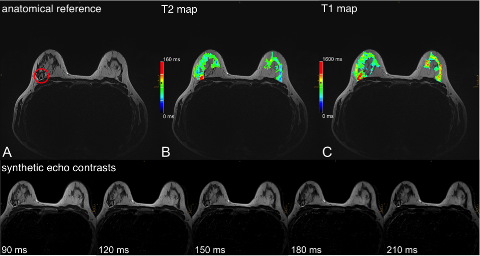
Figure 3 – 35-year-old patient with right-sided breast cancer with good response
after neoadjuvant chemotherapy showing a mostly necrotic tumor; A –
transversal T2w-TSE with a red circle marking the tumor; B – T2w-TSE with the T2 map from GRAPPATINI
as color-coded overlay with C/W windowing set to 80/160 ms; C – T2w-TSE with
the T1 map from the compressed sensing MP2RAGE prototype as color-coded overlay
with C/W 80/160 ms. The lower row illustrates the synthetic morphological
images derived from GRAPPATINI at effective TE of 90, 120, 150, 180, 210 ms
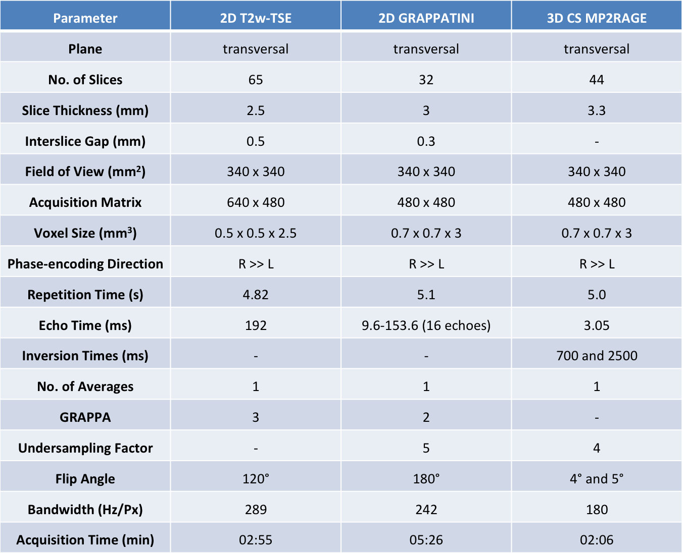
Table 1 – Overview of all parameters for the conventional T2-weighted (T2w)
turbo spin echo (TSE) sequence and the prototype T2 mapping (GRAPPATINI) and T1
mapping (compressed sensing MP2RAGE) sequence used in this study.