Nathan Tibbitts Roberts1,2, Diego Hernando1,2,3,4, Timothy Colgan1, Daiki Tamada1, and Scott B Reeder1,3,4,5,6
1Radiology, University of Wisconsin - Madison, Madison, WI, United States, 2Electrical and Computer Engineering, University of Wisconsin - Madison, Madison, WI, United States, 3Medical Physics, University of Wisconsin - Madison, Madison, WI, United States, 4Biomedical Engineering, University of Wisconsin - Madison, Madison, WI, United States, 5Medicine, University of Wisconsin - Madison, Madison, WI, United States, 6Emergency Medicine, University of Wisconsin - Madison, Madison, WI, United States
1Radiology, University of Wisconsin - Madison, Madison, WI, United States, 2Electrical and Computer Engineering, University of Wisconsin - Madison, Madison, WI, United States, 3Medical Physics, University of Wisconsin - Madison, Madison, WI, United States, 4Biomedical Engineering, University of Wisconsin - Madison, Madison, WI, United States, 5Medicine, University of Wisconsin - Madison, Madison, WI, United States, 6Emergency Medicine, University of Wisconsin - Madison, Madison, WI, United States
This work describes the successful development a novel B1- and fat-corrected CSE-MRI T1 mapping method. The proposed strategy demonstrates initial feasibility for a rapid, breath hold eligible, B1- and fat-corrected T1-mapping technique without the need for a separate B1 calibration scan.
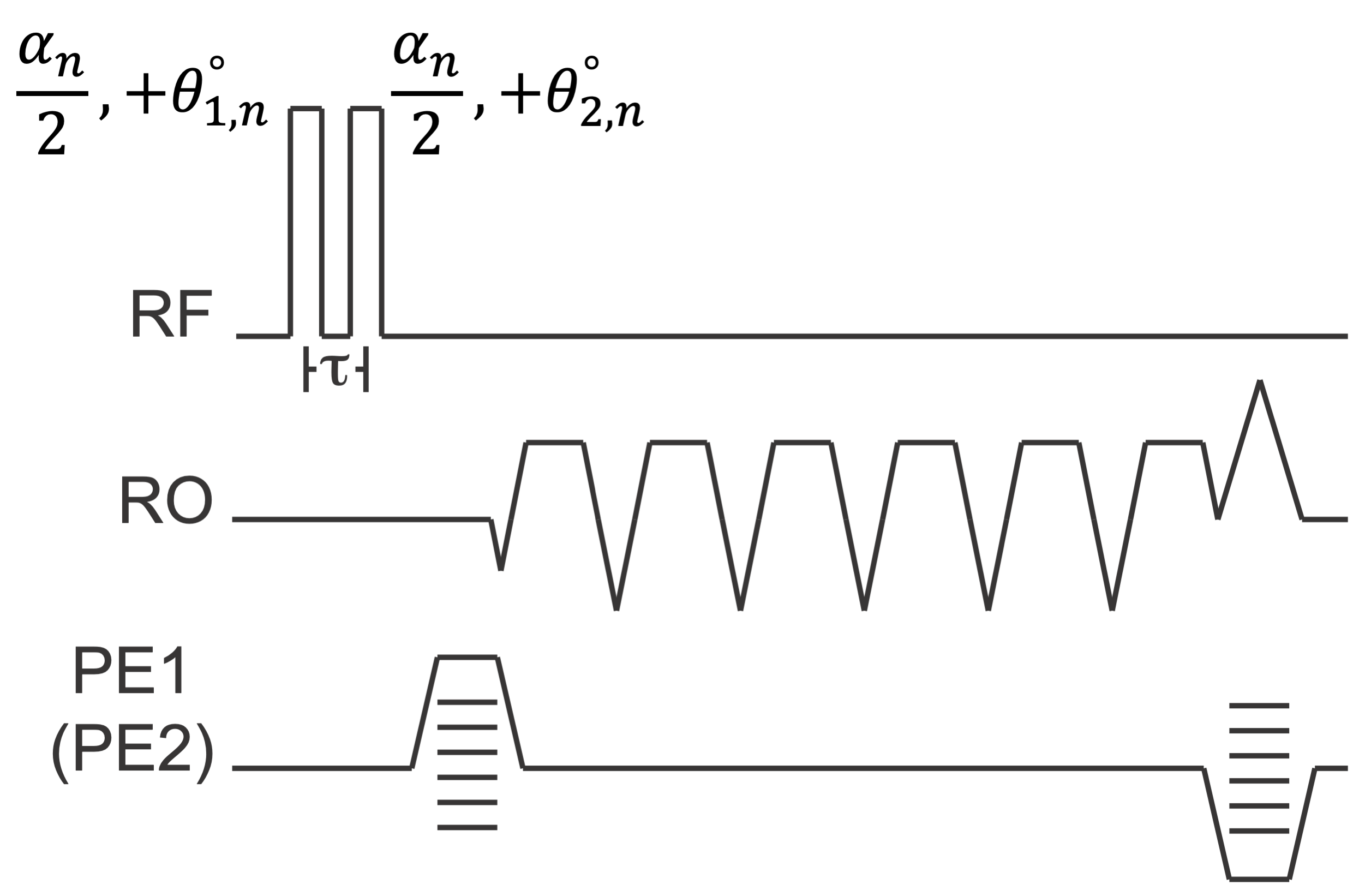
Figure 1. The pulse sequence of the proposed method is a two-pass (n=1,2) multi-echo 3D-SGRE sequence with a modified composite RF excitation module. Excitation phases are selected to ensure that the two RF pulses are orthogonal (θ1,1 = 0o, θ2,1 = 90o, θ1,2 = 90o, θ2,2 = 0o). Additionally, the order of RF pulses is toggled in the seconds pass to facilitate the elimination of background phase.
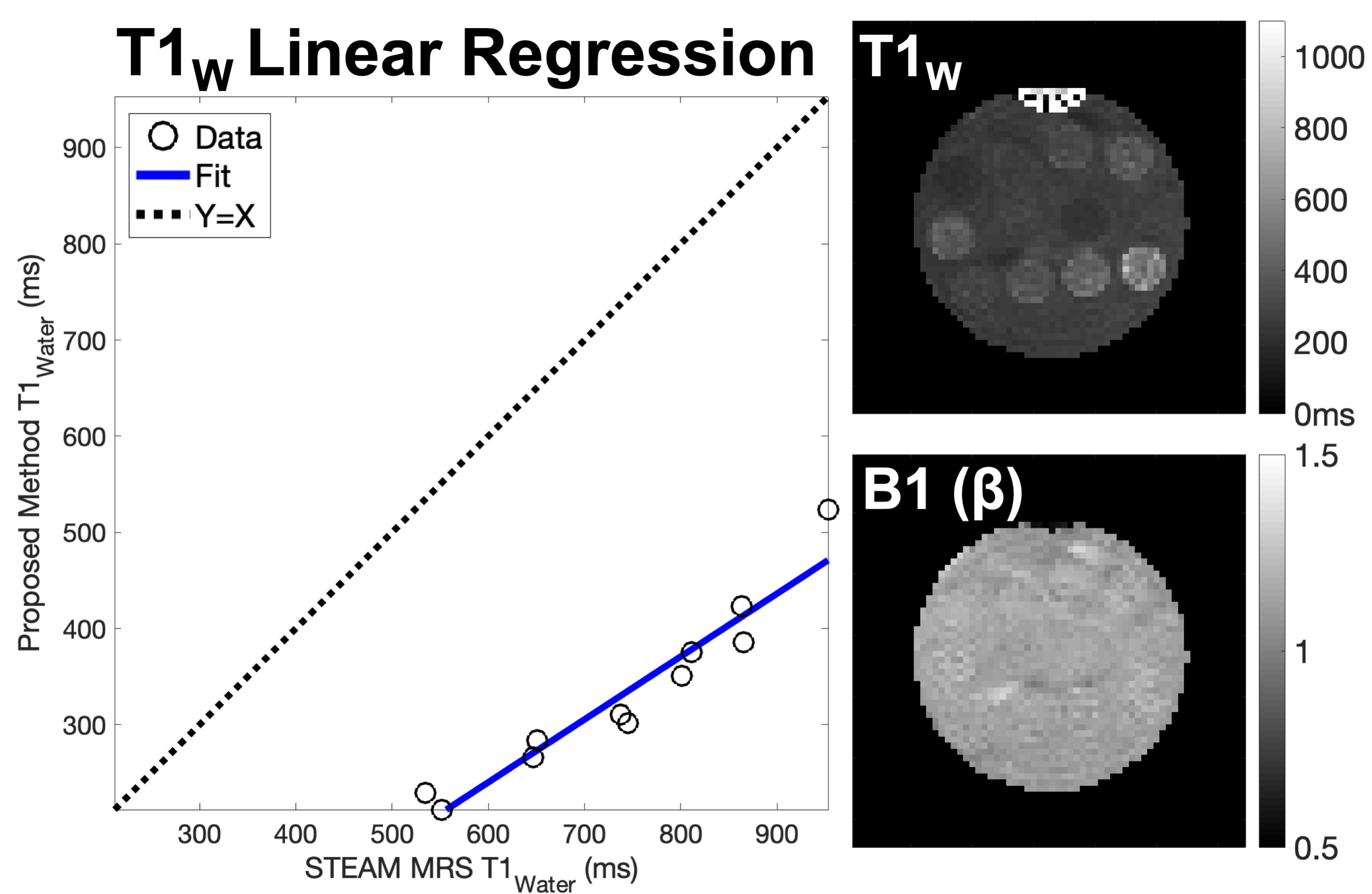
Figure 4. Estimates of T1W measured with the proposed method were highly correlated with the reference STEAM T1W, but also exhibited bias. Reconstructed T1W and B1 (β) are shown above from images acquired with a single channel quadrature head coil on a 1.5T system (GE Healthcare Optima MR450W, Waukesha, WI) with the following protocol specific parameters: Flip Angles: α1=50o, α2=30o, TR1=11.6ms, TR2=11.6ms, TE1=1.08ms, ΔTE=1.7ms, Echo ETL1=6, ETL2=6. Linear regression compares estimated T1W values to reference STEAM measurements in each vial.