Ya-Fang Chen1, Sung-Chun Tang2, and Wen-Chau Wu3,4
1Department of Medical Imaging, National Taiwan University Hospital Hsin-Chu Branch, Hsinchu, Taiwan, 2Department of Neurology, National Taiwan University Hospital, Taipei, Taiwan, 3Institute of Medical Device and Imaging, National Taiwan University, Taipei, Taiwan, 4Graduate Institute of Clinical Medicine, National Taiwan University, Taipei, Taiwan
1Department of Medical Imaging, National Taiwan University Hospital Hsin-Chu Branch, Hsinchu, Taiwan, 2Department of Neurology, National Taiwan University Hospital, Taipei, Taiwan, 3Institute of Medical Device and Imaging, National Taiwan University, Taipei, Taiwan, 4Graduate Institute of Clinical Medicine, National Taiwan University, Taipei, Taiwan
The
CBF derived from multi-PLD ASL varies with the choice of PLD (overestimated
when the PLD range does not cover ATT) as well as SNR (overestimated along with
greater variability when SNR is low).
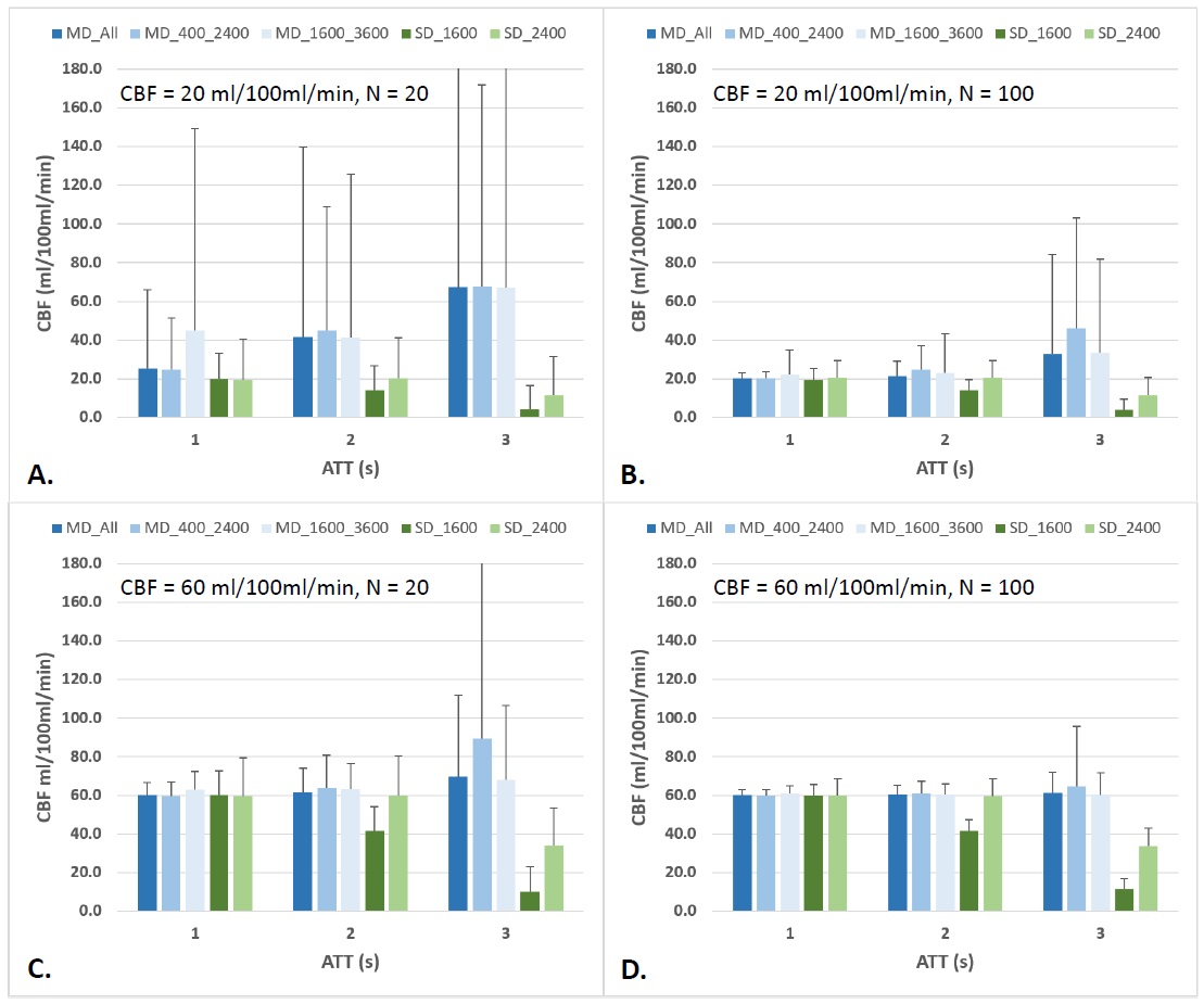
Figure
1. Simulated CBF measurement with multi-PLD and single-PLD methods. MD_400_2400
= multi-delay with 6 PLDs (400-2400 ms, in steps of 400 ms). MD_1600_3600 =
multi-delay with 6 PLDs (1600-3600 ms, in steps of 400 ms). MD_All =
multi-delay with 9 PLDs (400-3600 ms, in steps of 400 ms). SD_1600 =
single-delay with PLD = 1600 ms. SD_2400 = single-delay with PLD = 2400 ms. The
error bars indicate the standard deviation of 1000 calculations.
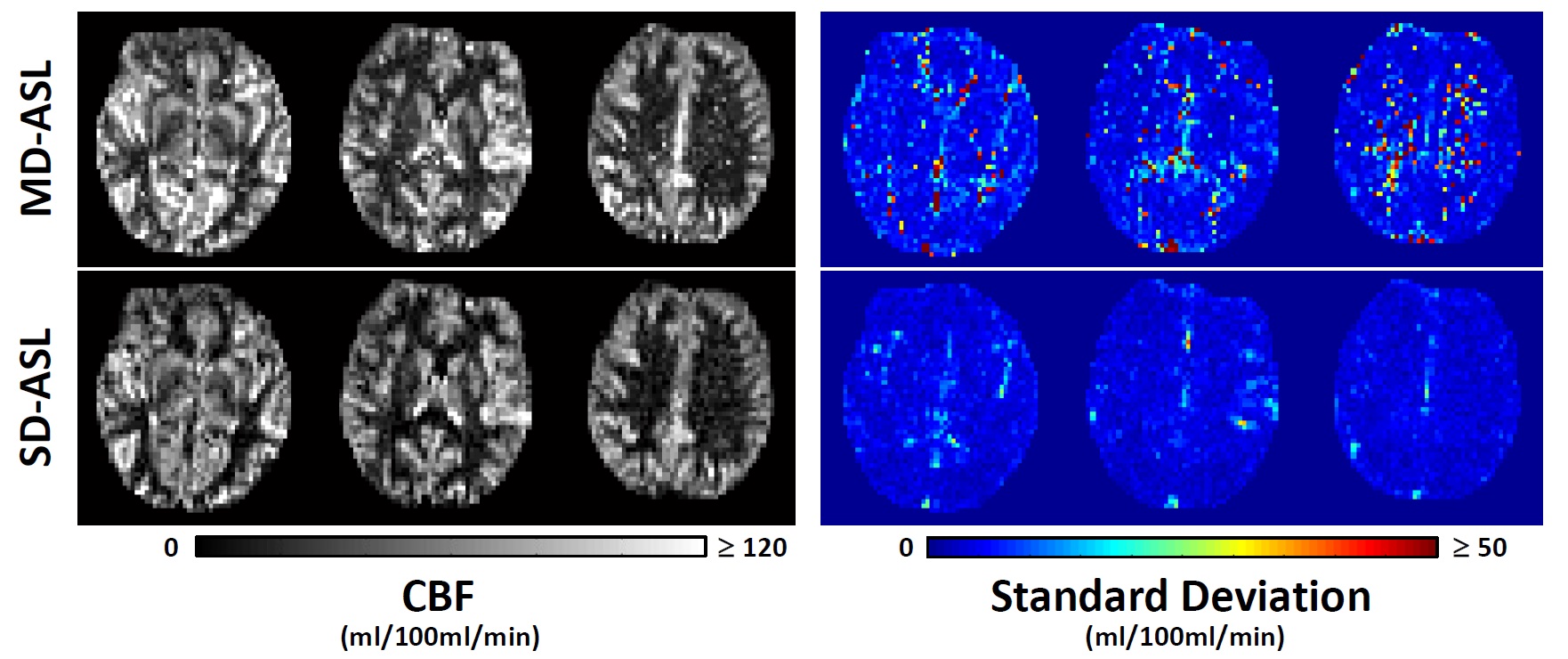
Figure
2. Representative maps of CBF (left panel) and standard deviation (right panel)
obtained from a healthy subject. MD = multi-delay. SD = single-delay. The
standard deviation maps of SD-ASL have been scaled with respect to averages comparable
with the total scan time of MD-ASL.