Ruoxun Zi1, Dan Zhu1,2, Wenbo Li2,3, and Qin Qin2,3
1Department of Biomedical Engineering, Johns Hopkins University School of Medicine, Baltimore, MD, United States, 2Russell H. Morgan Department of Radiology and Radiological Science, Johns Hopkins University School of Medicine, Baltimore, MD, United States, 3F.M. Kirby Research Center for Functional Brain Imaging, Kennedy Krieger Institute, Baltimore, MD, United States
1Department of Biomedical Engineering, Johns Hopkins University School of Medicine, Baltimore, MD, United States, 2Russell H. Morgan Department of Radiology and Radiological Science, Johns Hopkins University School of Medicine, Baltimore, MD, United States, 3F.M. Kirby Research Center for Functional Brain Imaging, Kennedy Krieger Institute, Baltimore, MD, United States
We presented a T2 prepared stack-of-spiral GRE pulse sequence and model-based reconstruction method with spatial sparsity regularization for T2 mapping of brain, which provided good performance with nRMSE of the whole brain T2 estimation equal to 8.2%±0.5% with an acceleration factor of 5.
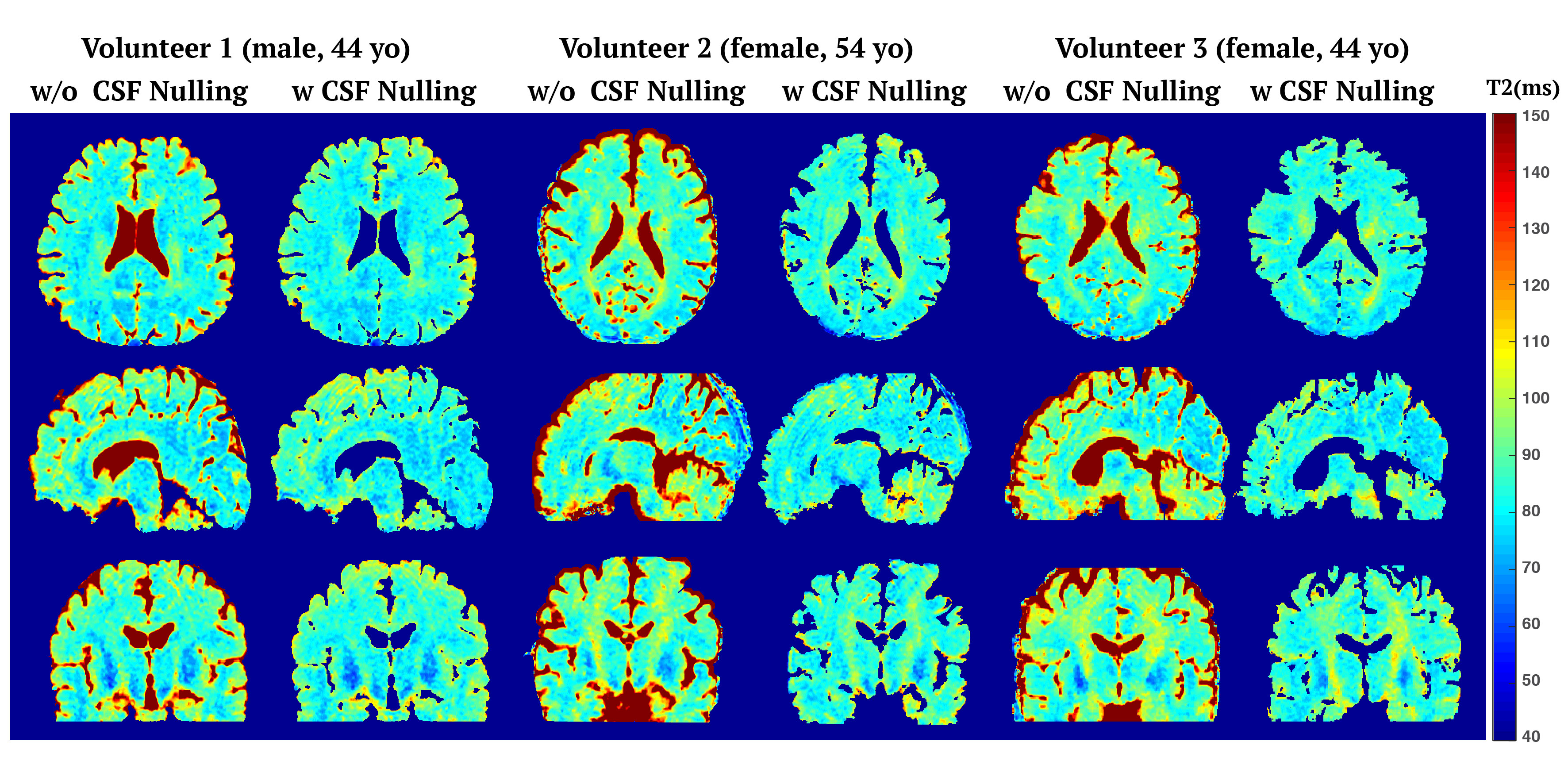
Figure 4. Cross-section T2 maps of brains of three healthy volunteers estimated with and without CSF nulling. In CSF nulling case, a mask was created based on the intensities of CSF-nulling images for better visualization and identification of lesion without CSF partial volume effect.
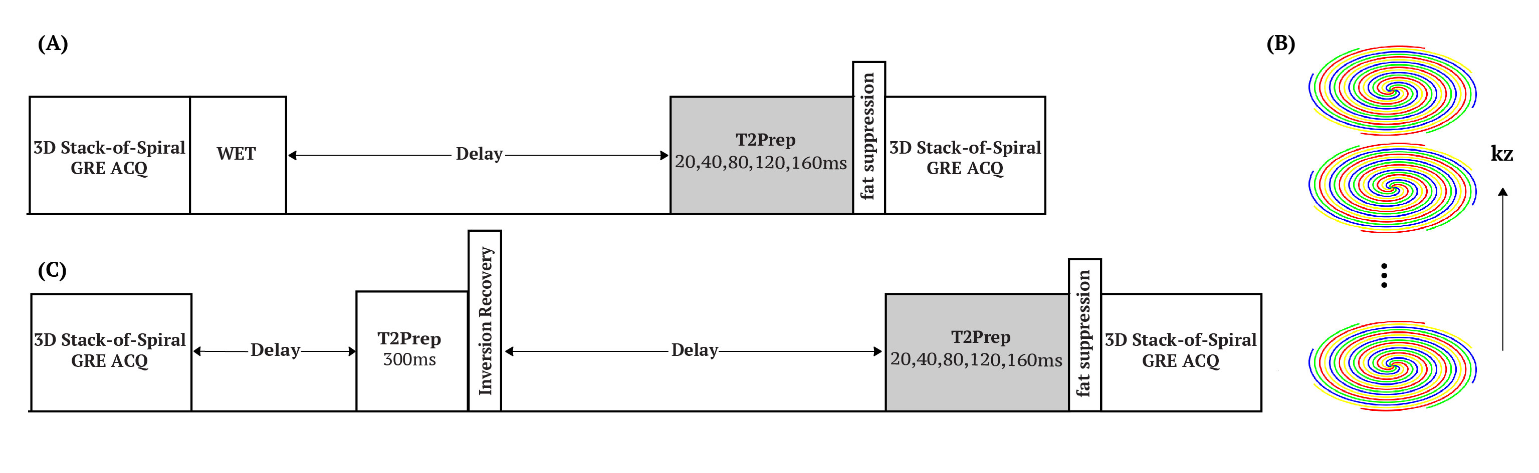
Figure 1. (A) Schematic diagram of T2 prepared 3D stack-of-spiral sequence. The duration of T2 preparation was set as 20,40,80,120,160ms. (B) 3D stack-of-spiral trajectory, with TFE encoding applied on kz dimension and variable density spiral trajectory (Accel=2) applied in plane. (C) the pulse sequence with CSF nulling to remove the CSF partial volume effect.
