Xinyuan Miao1,2, Yuankui Wu1,2,3, Dapeng Liu1,2, Qin Qin1,2, Peter C.M van Zijl1,2, Jay J. Pillai4,5, and Jun Hua1,2
1Neurosection, Division of MRI Research, Russell H. Morgan Department of Radiology and Radiological Science, Johns Hopkins University School of Medicine, Baltimore, MD, United States, 2F.M. Kirby Research Center for Functional Brain Imaging, Kennedy Krieger Institute, Baltimore, MD, United States, 3Department of Medical Imaging, Nanfang Hospital, Southern Medical University, Guangzhou, China, 4Johns Hopkins University School of Medicine, Division of Neuroradiology, Russell H. Morgan Department of Radiology and Radiological Science, Baltimore, MD, United States, 5Department of Neurosurgery, Johns Hopkins University School of Medicine, Baltimore, MD, United States
1Neurosection, Division of MRI Research, Russell H. Morgan Department of Radiology and Radiological Science, Johns Hopkins University School of Medicine, Baltimore, MD, United States, 2F.M. Kirby Research Center for Functional Brain Imaging, Kennedy Krieger Institute, Baltimore, MD, United States, 3Department of Medical Imaging, Nanfang Hospital, Southern Medical University, Guangzhou, China, 4Johns Hopkins University School of Medicine, Division of Neuroradiology, Russell H. Morgan Department of Radiology and Radiological Science, Baltimore, MD, United States, 5Department of Neurosurgery, Johns Hopkins University School of Medicine, Baltimore, MD, United States
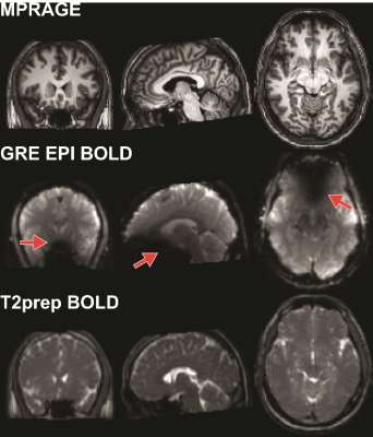
T2-prepared
blood oxygenation level–dependent functional MRI can significantly reduce
susceptibility artifacts commonly seen in gradient-echo echo-planar imaging functional
MRI in the presence of metallic orthodontic braces.

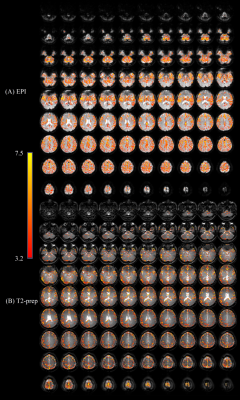
Figure 3: Activation maps overlaid on original fMRI images
from the GRE EPI (A) and T2prep (B) BOLD fMRI approaches, respectively from a
healthy subject wearing a titanium dental brace during a breath-hold task. The
activated voxels are highlighted with their corresponding t-scores from the GLM
analysis. The range of the t-scores is indicated by the scale bar.

