Maurizio Bergamino1, Lori Steffes1, Ashlyn Gonzales2, Leslie Baxter2, and Ashley M. Stokes1
1Neuroimaging Research, Barrow Neurological Institute, Phoenix, AZ, United States, 2Neuropsychology, Mayo Clinic Arizona, Phoenix, AZ, United States
1Neuroimaging Research, Barrow Neurological Institute, Phoenix, AZ, United States, 2Neuropsychology, Mayo Clinic Arizona, Phoenix, AZ, United States
SAGE-fMRI
with multiple echoes and contrast mechanisms was acquired and optimized for
fMRI analysis. SAGE-fMRI may ultimately provide new insight into the complex
contributions to BOLD signal and improve spatial localization of brain
activation.
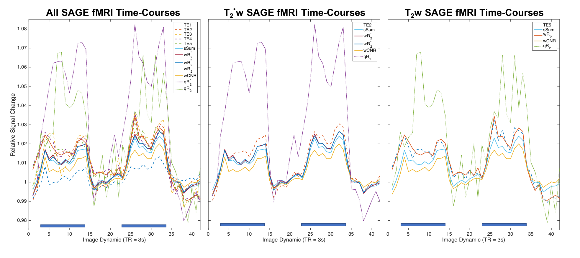
Dynamic fMRI data acquired using SAGE-EPI during a
visual task (“on” indicated by blue bar), normalized to 1 in the “off” dynamics,
in a 4x4x4 voxel ROI placed in the right occipital lobe. Left: All data
combinations (dashed lines: TE1-TE5; solid lines: multi-echo combinations;
dotted lines: quantitative R2* and R2). Middle and right panels show a subset
of the time-courses for T2*-weighting and T2-weighting, respectively. qR2* and qR2 have higher relative signal changes,
while TE1 has the smallest signal change.
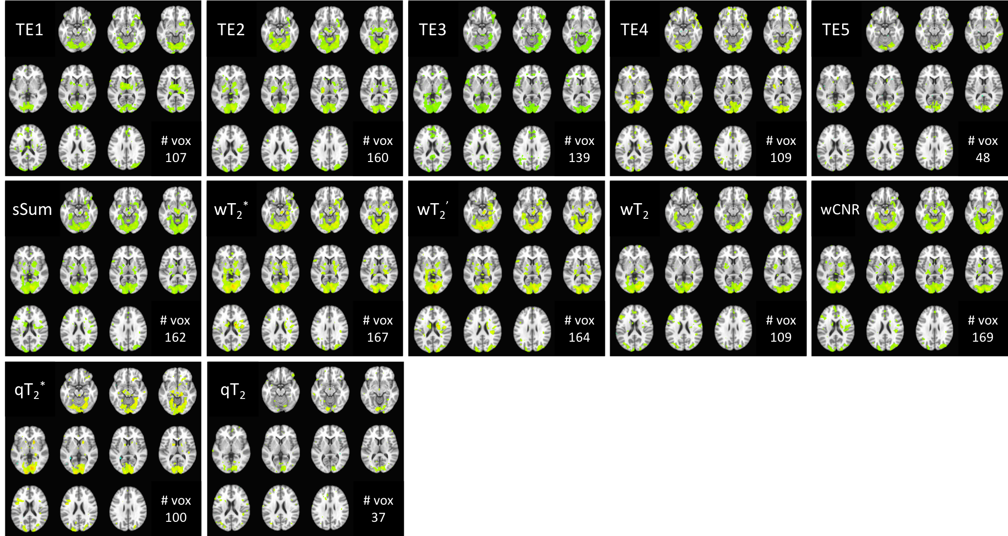
Group results for visual task SAGE-fMRI across single-echo (TE1-5, top row) and multi-echo combinations (weighted sums,
middle row; quantitative fits, bottom row). The number of significant voxels
(/1000) is indicated for each method at p<0.01. Of single-echo
analyses, TE2 has the highest number of voxels, but all weighted sum
combinations, except wT2, had more significant voxels. Relative
to TE5, weighting by T2 provides more voxels. Quantitative T2* and T2 yielded the smallest numbers of voxels per contrast mechanism, possibly reflecting higher accuracy for these metrics.