Luca Vizioli1, Steen Moeller1, Edward Auerbach1, Kamil Ugurbil1, and Essa Yacoub1
1CMRR, University of Minnesota, Minneapolis, MN, United States
1CMRR, University of Minnesota, Minneapolis, MN, United States
We show that by using a
multislab multiband 3D EPI approach we can achieve simultaneously high spatial
and temporal resolution for sub-millimeter fMRI applications at 7T.

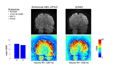
Example
of a single volume 3D GE (top left) and 2D GE (top right) EPI. Sequence details
are as follows: 3D GE-EPI: isotropic 0.8 mm3, TE = 23.4 ms, flip angle = 20°, 40 slices, TR =
143 ms, VAT = 1287 ms, iPAT = 3, partial Fourier = 6/8, MB = 2, z partial
fourier = 6/8; 2D GE-EPI:
isotropic 0.8 mm3, TE = 25.8 ms, flip angle = 57°, 40 slices, TR
= 1236 ms, iPAT = 3, partial Fourier = 6/8,
MB = 2).
The
bottom row shows tSNR maps. The blue bar plots represent the mean tSNR across
all voxels within the brain (including cerebellum). Errorbars reflect standard
error across voxels.

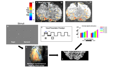
A) Average beta maps elicited by target and surround
for 3D and 2D GE EPI. B) Visual stimuli used. C) Visual block design paradigm.
D) Retinotopic representation of target in V1 computed by subtracting the activity
elicited by the target to that elicited by the surround. E) Equivolume cortical
depths in right V1. F) Activation for each cortical depth elicited by the target
condition within the retinotopic representation of the target in right V1. We
used independent data sets to define our ROI and extract the activation of each
depth. Errorbars show the standard errors across voxels.