Kirsty Hett1, Eleonora Patitucci1, Hannah Chandler1, Michael Germuska1, Benjamin Hope-Gill2, and Richard Wise1,3
1Cardiff University Brain Research Imaging Centre (CUBRIC), Cardiff University, Cardiff, United Kingdom, 2Respiratory Medicine, Cardiff and Vale University Health Board, Cardiff, United Kingdom, 3Institute for Advanced Biomedical Technologies, Department of Neuroscience, Imaging and Clinical Sciences, "G. D'Annunzio University" of Chieti-Pescara, Chieti, Italy
1Cardiff University Brain Research Imaging Centre (CUBRIC), Cardiff University, Cardiff, United Kingdom, 2Respiratory Medicine, Cardiff and Vale University Health Board, Cardiff, United Kingdom, 3Institute for Advanced Biomedical Technologies, Department of Neuroscience, Imaging and Clinical Sciences, "G. D'Annunzio University" of Chieti-Pescara, Chieti, Italy
A reduction in resting perfusion was seen in patients with Idiopathic Pulmonary Fibrosis (IPF) compared to matched controls. This was localised to the precuneus cortex, cingulate gyrus and lingual gyrus. There were no significant differences in grey matter volumes or functional response when performing a combined motor and visual task. Brain MRI was well tolerated in IPF patients supporting further investigation the cough pathway in IPF, which is a common and troubling symptom that is difficult to treat.
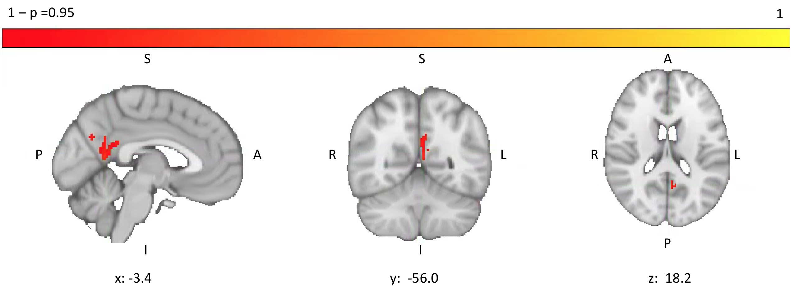
Figure 3: Areas of significantly higher perfusion in controls compared to patients with IPF in precuneous cortex, cingulate gyrus and lingual gyrus
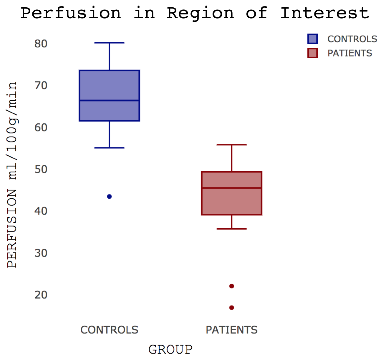
Figure 4: Boxplot showing differences in localised perfusion (precuneous cortex, cingulate gyrus and lingual gyrus, Figure 3) in controls and patients with IPF
A line across the box depicts the median, the box indicates the 25% and 75% percentiles, whiskers represent the maximum and minimum values, circles represent outliers.
A line across the box depicts the median, the box indicates the 25% and 75% percentiles, whiskers represent the maximum and minimum values, circles represent outliers.