Xiaoqing Alice Zhou1, Zengmin Li2, Hsu-Lei Lee3, Elizabeth Coulson4, and Kai-Hsiang Chuang3
1QBI/SBMS, The University of Queensland, Brisbane, Australia, 2QBI, The University of Queensland, Brisbane, Australia, 3QBI/CAI, The University of Queensland, Brisbane, Australia, 4SBMS/QBI, The University of Queensland, Brisbane, Australia
1QBI/SBMS, The University of Queensland, Brisbane, Australia, 2QBI, The University of Queensland, Brisbane, Australia, 3QBI/CAI, The University of Queensland, Brisbane, Australia, 4SBMS/QBI, The University of Queensland, Brisbane, Australia
BOLD activation by 1.5s short event can be reliably measured with ultrafast multiband EPI.
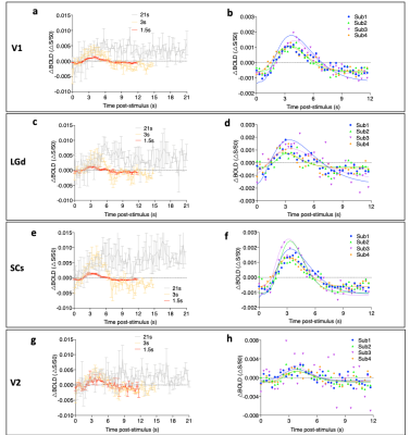
HRF in different brain regions of the visual pathway can be derived.
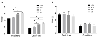
The HRF timing discrepancy may suggest timing difference in neural activity.

Fig.3 Temporal dynamics and HRF of evoked BOLD. a) Mean BOLD timecourse in response to 21/3/1.5s stimulations in the V1 (n=4). b)HRF of V1 modelled from a single gamma-variate function. c) Mean BOLD timecourse in response to 21/3/1.5s stimulation in the LGd (n=4). d) HRF of LGd. e) Mean BOLD timecourse in response to 21/3/1.5s stimulation in the SCs (n=4). f) HRF of SCs. g) Mean BOLD timecourse in response to 21/3/1.5s stimulation in the V2 (n=4). h) HRF of V2. Data are shown as mean±SEM.

