Yayan Yin1, Lang Qin2, and Jie Lu1
1Radiology, Capital Medical University, Beijing, China, 2Linguistics, the University of Hong Kong, Beijing, China
1Radiology, Capital Medical University, Beijing, China, 2Linguistics, the University of Hong Kong, Beijing, China
The present study is the first time to show the time course of OEF response for motor task in both normoxia and mild hypoxia. The results showed that mild hypoxia caused a larger OEF response during mild hypoxia and was positively correlated with oxygen saturation.
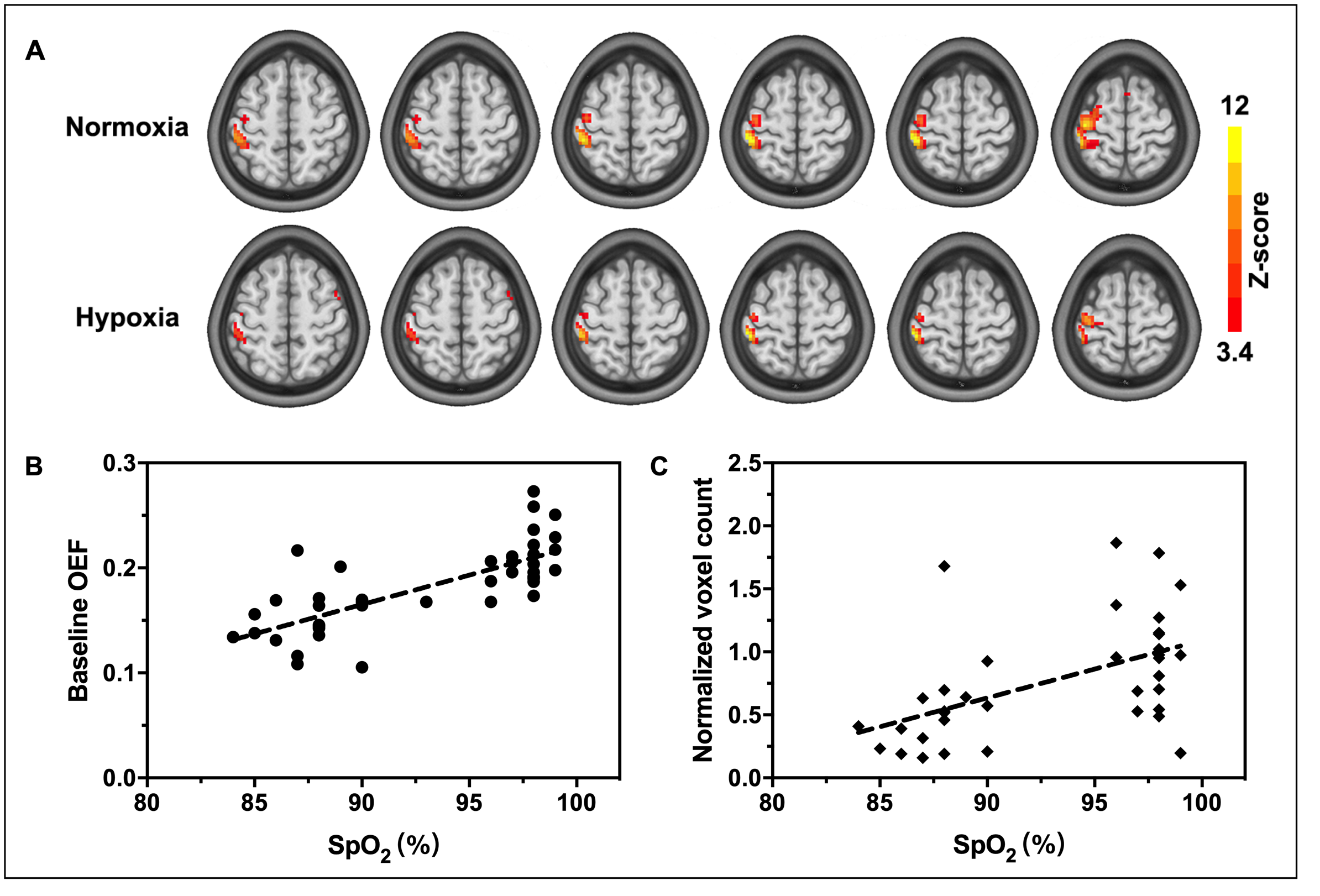
Figure 1. (A) OEF activation maps of a typical subject during a 1 Hz right hand grasp-release task for each gas condition. The activation region (in color) was generated using a voxel-level threshold of P< 0.001 with a cluster-level FWE correction of P< 0.05 superimposed onto the standard template. The baseline OEF signal (B) and the active voxels (C) were decreased with decreasing %SpO2.
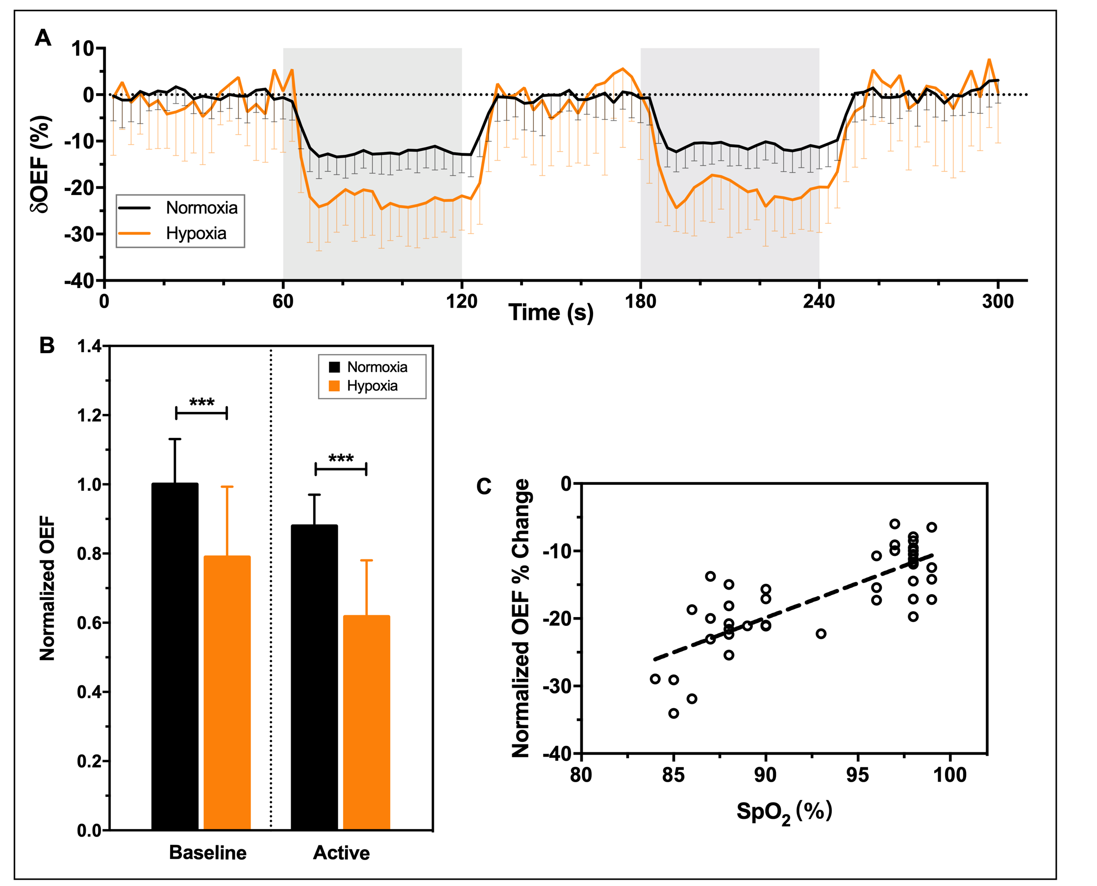
Figure 2. (A) Time courses of OEF signal from the overlapped voxels were normalized with its baseline OEF value for each gas condition. Data shown as mean±SD (n=21). The areas in gray represent periods of stimulation. (B) Normalized OEF in normoxia and hypoxia during baseline and activation (P< 0.0001). (C) Normalized OEF signal changes (P< 0.0001) were decreased with decreasing %SpO2.