Alexander M Puckett1, Zoey Isherwood2, Ashley York1, Catherine Viengkham3, and Branka Spehar3
1University of Queensland, Brisbane, Australia, 2University of Wollongong, Wollongong, Australia, 3University of New South Wales, Sydney, Australia
1University of Queensland, Brisbane, Australia, 2University of Wollongong, Wollongong, Australia, 3University of New South Wales, Sydney, Australia
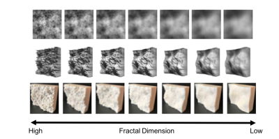
3D-printed
fractal textures can be used to elicit robust BOLD activation throughout human
primary somatosensory cortex, and fMRI at 7T can be used to measure this
activity within individual fingertip representations.

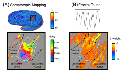
Figure 3. Somatotopic fingertip mapping and the fractal touch
experiment elicit overlapping activity. (A) Somatotopic
mapping data projected onto an inflated cortical surface model. Top shows an un-thresholded activation map.
Bottom shows the fingertip map within a zoomed in area around the post-central
gyrus. White dashed line illustrates the S1 ROI boundary. (B) Bottom shows the activation
map in and near S1 during the fractal touch experiment, thresholded at a
FDR-corrected q < 0.001. An example of a single voxel time-course is shown
above.
