Yixin Ma1,2, Julia Bresticker1,2, Dean Darnell1,2, Fraser Robb3, Allen Song1,2, and Trong-Kha Truong1,2
1Brain Imaging and Analysis Center, Duke University, Durham, NC, United States, 2Medical Physics Graduate Program, Duke University, Durham, NC, United States, 3GE Healthcare, Aurora, OH, United States
1Brain Imaging and Analysis Center, Duke University, Durham, NC, United States, 2Medical Physics Graduate Program, Duke University, Durham, NC, United States, 3GE Healthcare, Aurora, OH, United States
In vivo experiments with an adaptive iPRES(2) body coil array demonstrated that it can reduce B0 inhomogeneities and EPI distortions more effectively than an iPRES(1) coil array, without requiring additional power supplies.
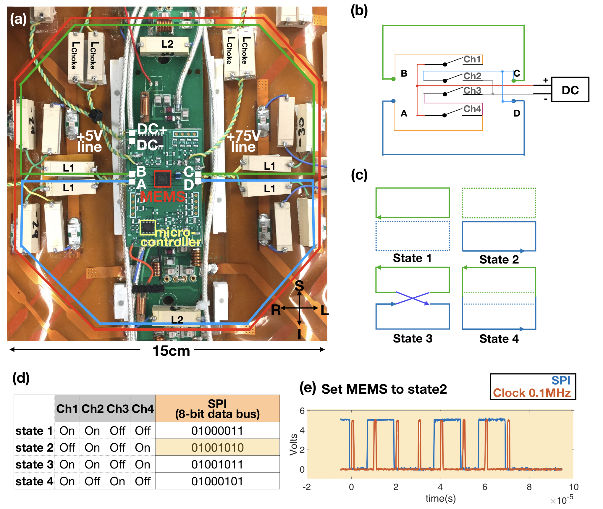
Fig. 1. (a) iPRES(2)-A coil element with: interior DC traces, inductors (L1/L2), DC power supply line (DC±) with chokes (Lchoke), MEMS switch package, 75-V and 5-V bias supply lines, and micro-controller. (b) Diagram of a MEMS switch controlling the DC current flow between two iPRES(2)-A shim loops (blue/green). (c) Four iPRES(2)-A states, and (d) configurations of MEMS switch channels. (e) 8-bit serial logical command and clock lines to set the MEMS switch state.
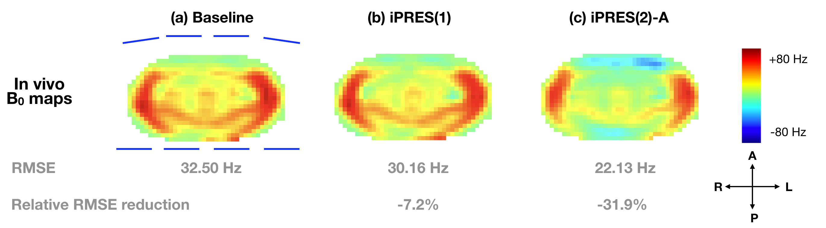
Fig. 3. B0 maps (Hz) and RMSEs in a representative slice before (a) and after iPRES(2)-A (b) or iPRES(1) (c) shimming. The B0 maps were acquired with a multi-echo gradient-echo sequence and reconstructed with a multipoint Dixon fat-water separation method5. The blue lines surrounding the baseline B0 map indicate the positions of the eight iPRES(2)-A coil elements relative to the subject. iPRES(2)-A can greatly reduce localized B0 inhomogeneities compared to the baseline or iPRES(1).