Jan Morez1, Jan Sijbers1, and Ben Jeurissen1
1imec-Vision Lab, Dept. Physics, University of Antwerp, Antwerp, Belgium
1imec-Vision Lab, Dept. Physics, University of Antwerp, Antwerp, Belgium
We present a 5-minute scanning protocol for multi-tissue constrained spherical deconvolution, optimized for precise WM and GM density estimations, while maintaining a high angular resolution.
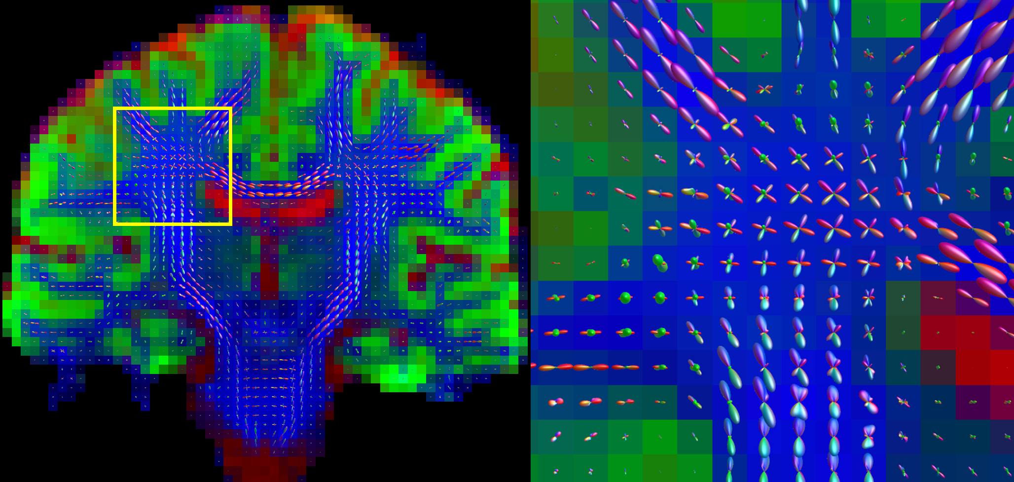
Figure 3, left) tissue densities (red: CSF, green: GM, blue: WM) and WM ODFs estimated from a data set acquired with the optimal sampling. right) close-up of the WM ODFs delineated by the yellow square.

Figure 2: a) WM, GM and CSF density variance maps calculated from the real data acquired with the reference and optimized DW schemes. b) distribution of variances depicted in (a). The median variances of these maps are for the reference and optimized schemes, respectively: 5.58e-4 and 3.74e-4 (WM), 5.54e-4 and 4.38e-4 (GM), 5.00e-5 and 7.94e-5 (CSF).