Lauren Scott1, Ben Dickie1, Damien McHugh2, John McFadden1, Andrew N Priest3, and Laura M Parkes1
1Division of Neuroscience and Experimental Psychology, University of Manchester, Manchester, United Kingdom, 2Quantitative Biomedical Imaging Laboratory, University of Manchester, Manchester, United Kingdom, 3Radiology, Cambridge University Hospitals NHS Foundation Trust, Cambridge, United Kingdom
1Division of Neuroscience and Experimental Psychology, University of Manchester, Manchester, United Kingdom, 2Quantitative Biomedical Imaging Laboratory, University of Manchester, Manchester, United Kingdom, 3Radiology, Cambridge University Hospitals NHS Foundation Trust, Cambridge, United Kingdom
Varying
diffusion-time introduces sensitivity to microvascular blood velocity, structure,
and transvascular water exchange. Parameter estimates consistent with
literature values are found in the cerebral cortex and white matter of healthy
volunteers.
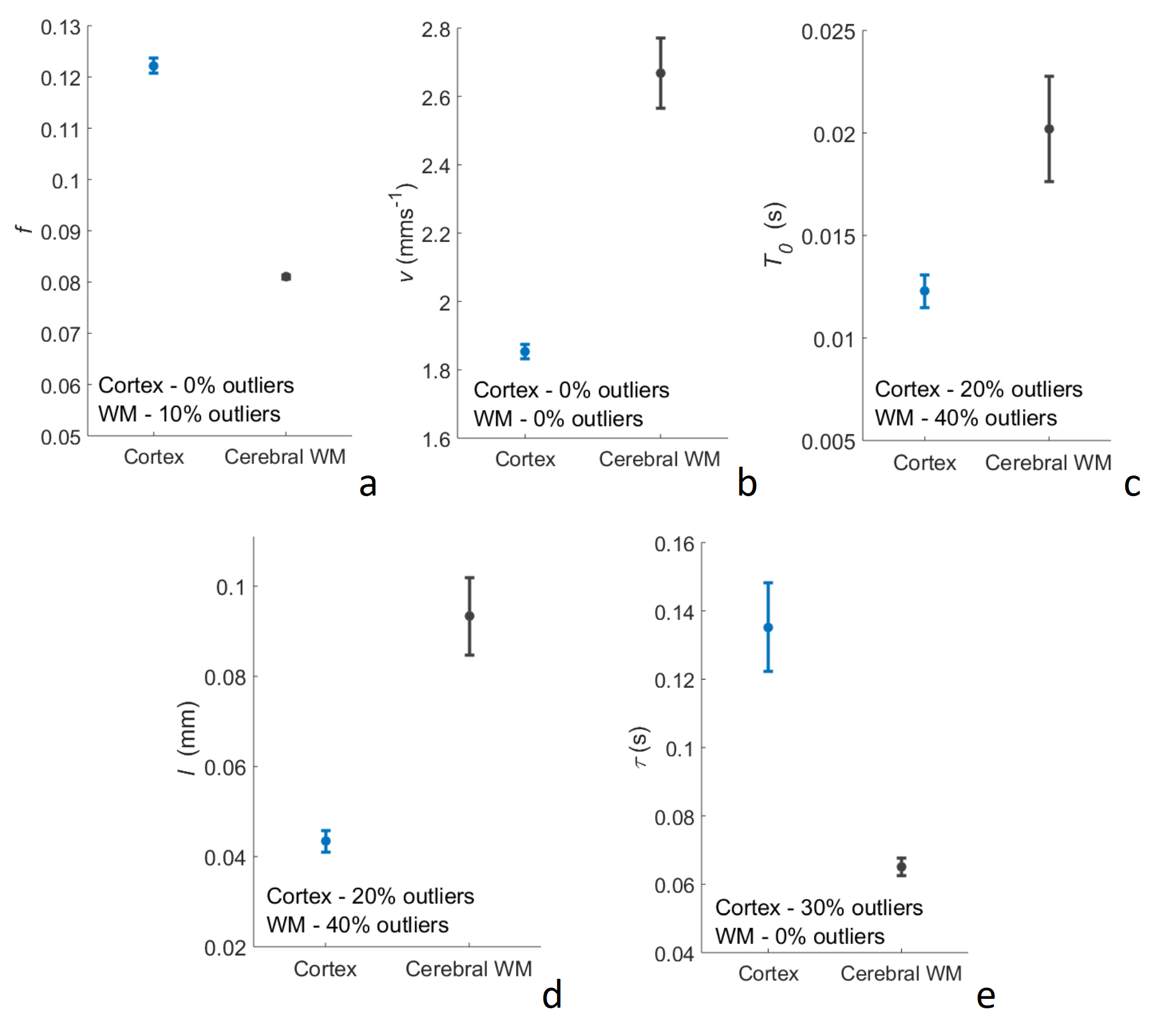
Figure 2: Parameter
estimates from a two-step fit to DW-MR data. Mean parameter estimates ± SE are shown over 10 participants
for the cerebral cortex and white matter (WM). (a) shows the perfusion fraction, f; (b) the average blood flow velocity,
v; (c) the correlation time, T0;
(d) the average capillary segment length, l;
and (e) the mean water residence time in the intravascular compartment, τ.
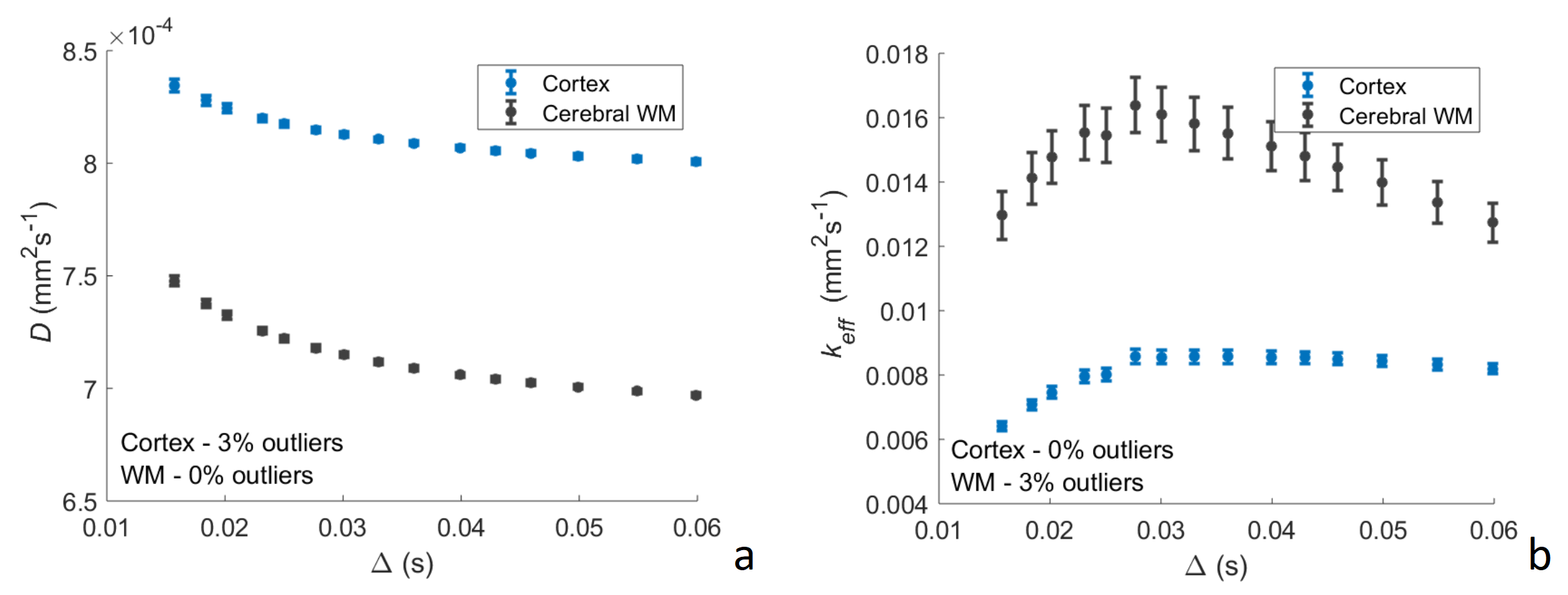
Figure 1: Diffusion-time
(∆) dependence of the diffusion (D) (a) and perfusion (keff) (b) coefficients in the cerebral cortex and white matter (WM). Mean parameter estimates ± SE
are shown over 10 participants. D
reduces with ∆ as the water accesses more barriers to diffusion. keff
behaves differently, initially increasing with ∆ as greater distances are reached, and plateauing
as water begins to change direction in the isotropic capillary bed. keff falls at longer ∆ as
the perfusing component exchanges with the less strongly dephased extravascular
component.