Gregory Lemberskiy1,2, Yousef Mazaheri3, Herbert Alberto Vargas3, Ricardo Otazo3, Els Fieremans1, and Dmitry S Novikov1
1Radiology, New York University School of Medicine, New York, NY, United States, 2Microstructure Imaging INC, New York, NY, United States, 3Memorial Sloan Kettering Cancer Center, New York, NY, United States
1Radiology, New York University School of Medicine, New York, NY, United States, 2Microstructure Imaging INC, New York, NY, United States, 3Memorial Sloan Kettering Cancer Center, New York, NY, United States
3 prostate cancer patients with PIRADS≥4
lesions were images with a multi-echo/multi-diffusion time protocol. We find
that varying the echo time for prostate cancer diffusion weighted imaging increases ADC
for benign tissue while minimally
affecting the malignant tissue.
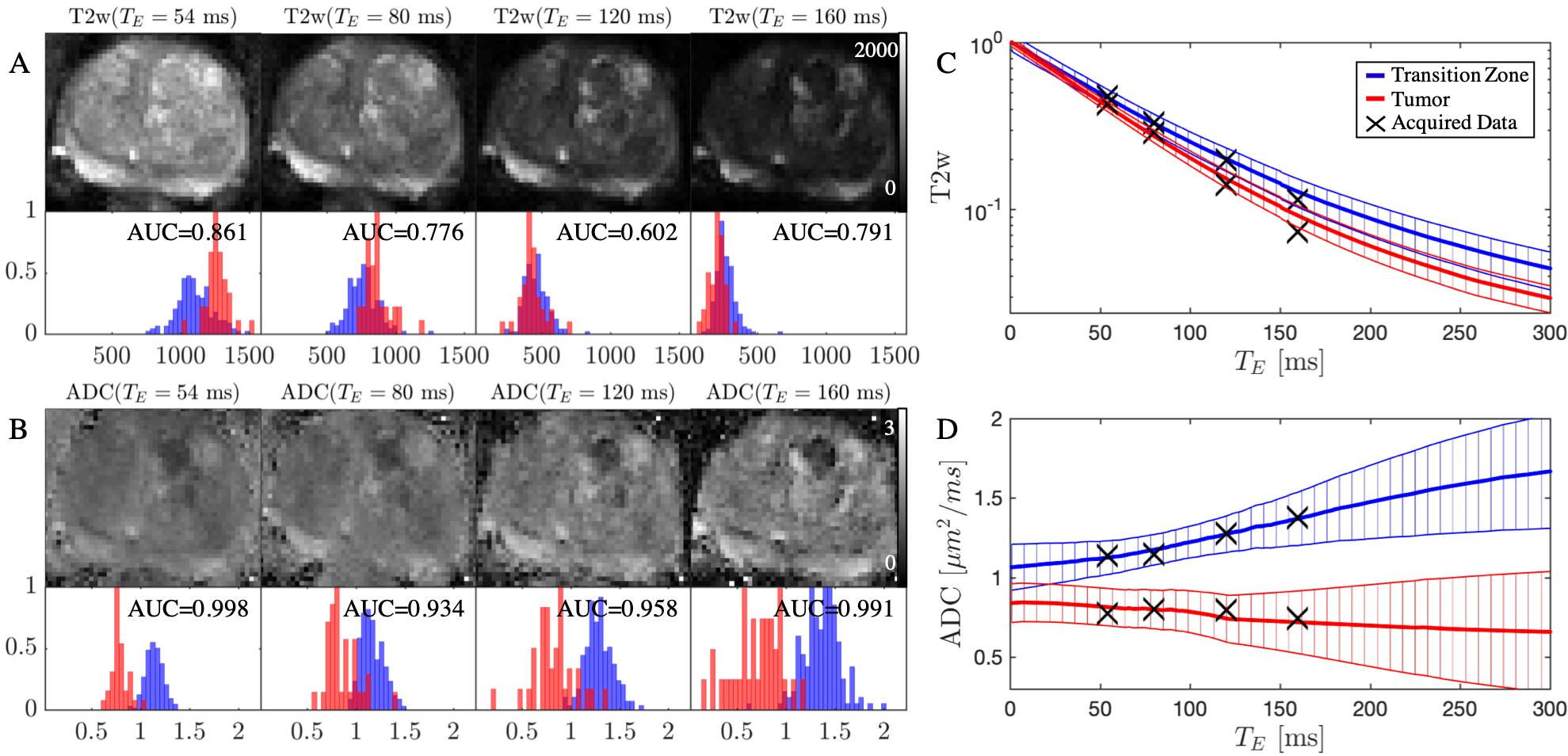
Figure 2: Echo time
dependence on $$$T_2w$$$ and $$$ADC$$$. (A) Diffusion-free $$$T_2w$$$ images $$$S|_{b=0}(T_E)$$$ and (B) $$$ADC$$$ maps for increasing $$$T_E$$$ for patient 1. For each
image, the corresponding histograms are shown for benign transition zone (blue)
and PIRADS-5 lesion (red). The mean and standard deviation of the $$$T_E$$$ dependence
on (C) $$$T_2w$$$ and (D) $$$ADC$$$ is extrapolated for benign and malignant transition
zone. At long $$$T_E$$$, the benign tissue $$$ADC$$$ increases, while the lesion $$$ADC$$$ is
nearly constant.
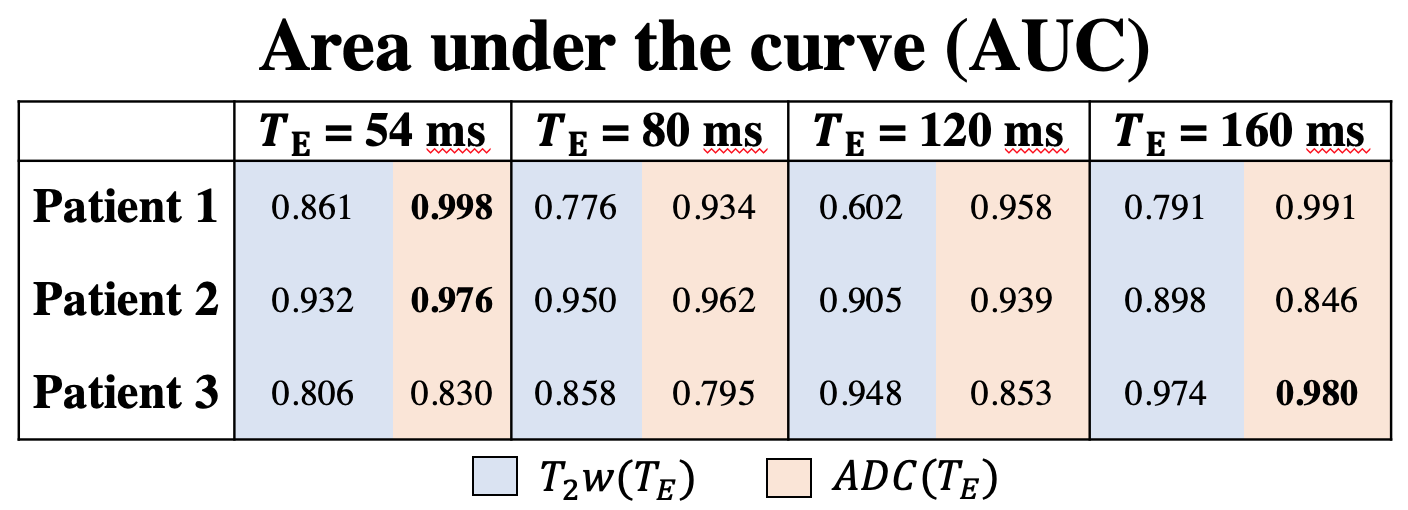
Figure 3: Area under the curve (AUC) plotted as
a function of TE, separating between benign and malignant transition zone in 3
patients.