Li Zhao1, Xue Feng2, Josepheen Asis-Cruz 1, Yao Wu1, Kushal Kapse1, Axel Ludwig1, Dan Wu3, Kun Qing2, Carig H. Meyer2, and Catherine Limperopoulos1
1Childrens National Hospital, Washington, DC, United States, 2University of Virginia, Charlottesville, VA, United States, 3Zhejiang University, Hanzhou, China
1Childrens National Hospital, Washington, DC, United States, 2University of Virginia, Charlottesville, VA, United States, 3Zhejiang University, Hanzhou, China
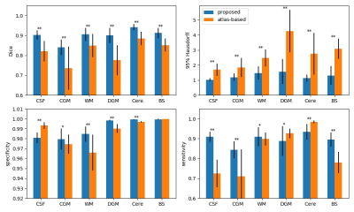
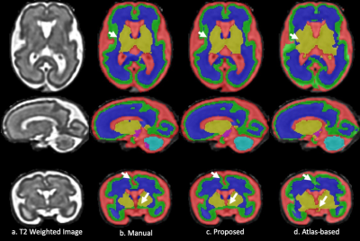
A fetal brain segmentation method was developed based on 3D U-Net, which
provided faster, higher accuracy segmentation and consistent performance across
gestational ages, compared to the conventional method based on atlases.


