Jeffry R. Alger1,2,3, Joseph O'Neill4, Lisa Kilpatrick4, Katherine L. Narr1, Guldamla Kalender4, Ronald Ly4, Shantanu H. Joshi1, Sandy Loo4, Mary J. O'Connor4, and Jennifer G. Levitt4
1Neurology, University of California, Los Angeles, Los Angeles, CA, United States, 2Advanced Imaging Research Center, University of Texas Southwestern Medical Center, Dallas, TX, United States, 3NeuroSpectroScopics LLC, Sherman Oaks, CA, United States, 4Psychiatry and Biobehavioral Sciences, University of California, Los Angeles, Los Angeles, CA, United States
1Neurology, University of California, Los Angeles, Los Angeles, CA, United States, 2Advanced Imaging Research Center, University of Texas Southwestern Medical Center, Dallas, TX, United States, 3NeuroSpectroScopics LLC, Sherman Oaks, CA, United States, 4Psychiatry and Biobehavioral Sciences, University of California, Los Angeles, Los Angeles, CA, United States
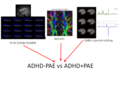
Multimodal MR
neuroimaging identified brain differences ADHD+PAE, ADHD-PAE and controls in an study of children aged 8-12 years. The findings provide
preliminary support for the hypothesis that an objective diagnostic neuroimaging-based
classifier can be developed.

