Ruohan Feng1, Lihua Zhuo1, Xinyue Hu2, Yingxue Gao2, Lianqing Zhang2, Yang Li3, Xinyu Hu2, Shi Tang2, Ming Zhou1, Guoping Huang3, and Xiaoqi Huang2
1Department of Radiology, the third hospital of Mianyang, Mianyang, China, Mianyang, China, 2Huaxi MR Research Center (HMRRC), Functional and Molecular Imaging Key Laboratory of Sichuan Province, Department of Radiology, West China Hospital, Sichuan University, Chengdu 610041, China, Chengdu, China, 3Department of Psychiatry, the third hospital of Mianyang, Mianyang, China, Mianyang, China
1Department of Radiology, the third hospital of Mianyang, Mianyang, China, Mianyang, China, 2Huaxi MR Research Center (HMRRC), Functional and Molecular Imaging Key Laboratory of Sichuan Province, Department of Radiology, West China Hospital, Sichuan University, Chengdu 610041, China, Chengdu, China, 3Department of Psychiatry, the third hospital of Mianyang, Mianyang, China, Mianyang, China
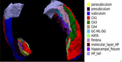
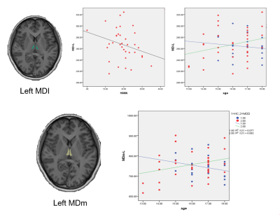
Compared to HCs, the MDD adolescents showed reduction in volume of subfields of the hippocampus, and increased volume in the subfileds of the thalamus, while no significant difference in amygdala subregions was found.

