Nathanael Kim1, Yousef Mazaheri1, Yulia Lakhman2, Li Feng3, Ersin Bayram4, Alberto Vargas2, and Ricardo Otazo1,2
1Medical Physics, Memorial Sloan Kettering Cancer Center, New York, NY, United States, 2Radiology, Memorial Sloan Kettering Cancer Center, New York, NY, United States, 3Biomedical Engineering and Imaging Institute and Department of Radiology, Icahn School of Medicine at Mount Sinai, New York, NY, United States, 4GE Healthcare, Waukesha, WI, United States
1Medical Physics, Memorial Sloan Kettering Cancer Center, New York, NY, United States, 2Radiology, Memorial Sloan Kettering Cancer Center, New York, NY, United States, 3Biomedical Engineering and Imaging Institute and Department of Radiology, Icahn School of Medicine at Mount Sinai, New York, NY, United States, 4GE Healthcare, Waukesha, WI, United States
In this work, we use the GRASP method to perform
DCE-MRI of gynecological tumors with high spatial and temporal resolution and to
estimate the AIF directly from the data. The personalized AIF shows higher
consistency with the tumor enhancement compared to the population AIF.
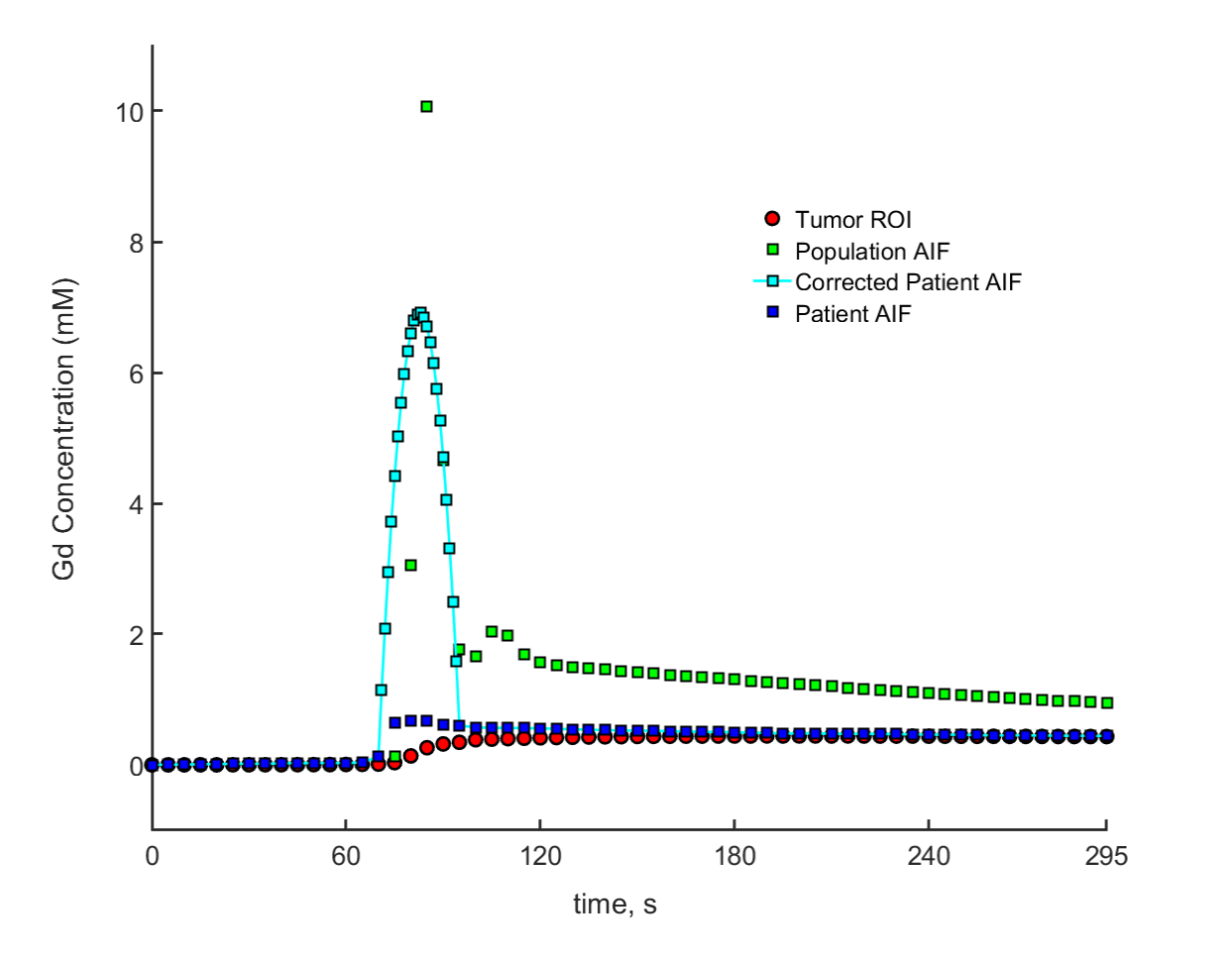
Figure 2: A graph showing the comparison of the
different AIFs, showing the correction made to the patient AIF by adjusting the
peak. The corrected patient AIF is more correlated with the tumor enhancement,
as expected from the personalized estimation.
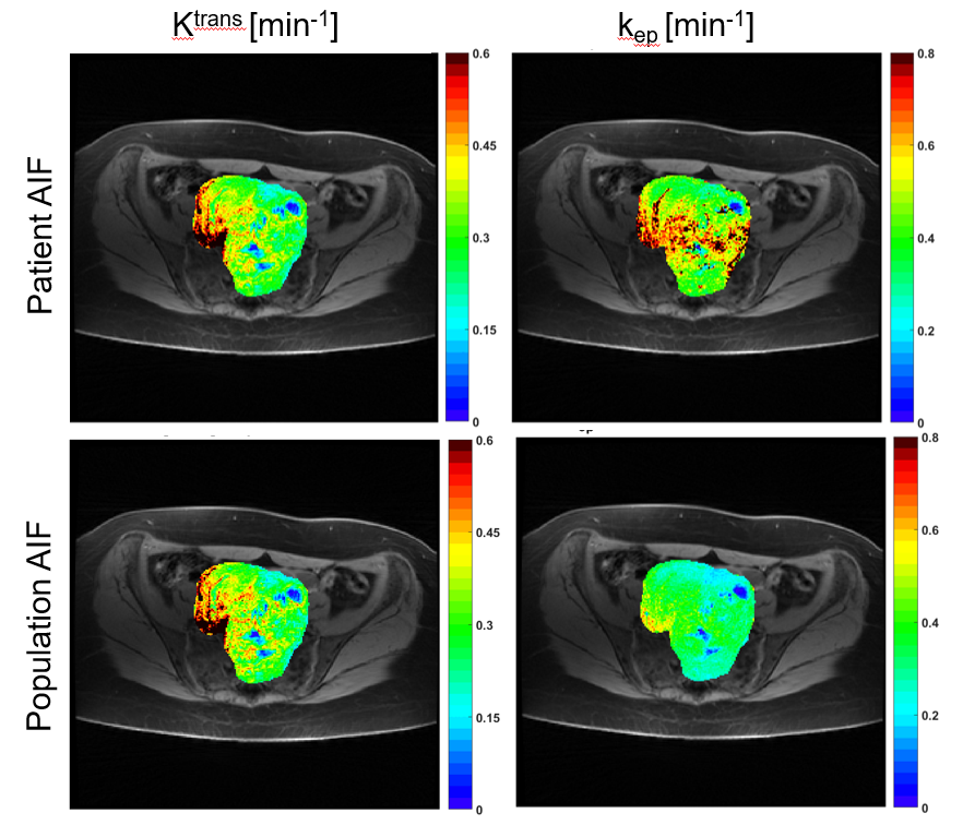
Figure 4: Another comparison of Ktrans and
kep maps for a patient with a large cervical tumor. As before, differences are more pronounced on
the kep map.