Digital Posters
Artifacts & Corrections Related to the Acquisition
ISMRM & SMRT Annual Meeting • 15-20 May 2021

Digital Posters
Artifacts & Corrections Related to the Acquisition
| Concurrent 1 | 13:00 - 14:00 |
3521.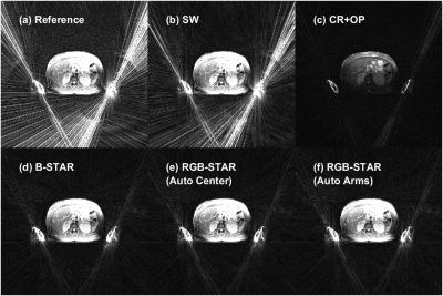 |
Automated Radial Streaking Artifact Suppression with RGB-STAR
Rohit Chacko Philip1, Ali Bilgin1,2,3, and Maria I Altbach1,2
1Medical Imaging, University of Arizona, Tucson, AZ, United States, 2Biomedical Engineering, University of Arizona, Tucson, AZ, United States, 3Electrical and Computer Engineering, University of Arizona, Tucson, AZ, United States
Streaking artifacts in radial MR imaging due to gradient nonlinearities are suppressed using a beamforming algorithm where region growing image segmentation is used to automatically generate the interference covariance matrix. The performance of the automatic streaking artifact suppression algorithm (RGB-STAR) is compared to algorithms based on coil removal and coil weighting and a beamforming algorithm with manual segmentation.
|
|||
3522.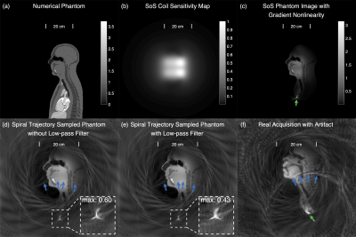 |
Aliasing Artifact Reduction in Spiral Real-Time MRI
Ye Tian1, Yongwan Lim1, Ziwei Zhao1, Dani Byrd2, Shrikanth Narayanan1,2, and Krishna S. Nayak1
1Ming Hsieh Department of Electrical and Computer Engineering, University of Southern California, Los Angeles, CA, United States, 2Department of Linguistics, University of Southern California, Los Angeles, CA, United States
Mid-sagittal spiral MRI of speech production often suffers from a distinct and disruptive aliasing artifact arising from a spurious signal outside the FOV. In this work, we determine that the spurious signal is caused by gradient nonlinearity and an ineffective anti-aliasing filter in spiral readout. We propose and evaluate two methods to mitigate the artifact, termed the large FOV (LF) method and the estimation-subtraction (ES) method. Qualitative evaluation score from two speech experts using a 5-level Likert scale improved 1.25 and 1.35 with 228.8% and 6.9% increment of reconstruction time for the LF and ES methods, respectively.
|
|||
3523.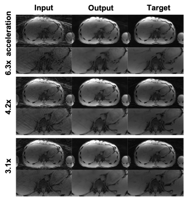 |
Streaking artifact reduction of free-breathing undersampled stack-of-radial MRI using a 3D generative adversarial network
Chang Gao1,2, Vahid Ghodrati1,2, Dylan Nguyen3, Marcel Dominik Nickel4, Thomas Vahle4, Brian Dale5, Xiaodong Zhong6, and Peng Hu1,2
1Department of Radiological Sciences, University of California Los Angeles, Los Angeles, CA, United States, 2Department of Physics and Biology in Medicine, University of California Los Angeles, Los Angeles, CA, United States, 3Department of Bioengineering, University of California Los Angeles, Los Angeles, CA, United States, 4MR Application Predevelopment, Siemens Healthcare GmbH, Erlangen, Germany, 5MR R&D Collaborations, Siemens Medical Solutions USA, Inc., Cary, NC, United States, 6MR R&D Collaborations, Siemens Medical Solutions USA, Inc., Los Angeles, CA, United States
Undersampling in free-breathing stack-of-radial MRI is desirable to shorten the scan time but introduces streaking artifacts. Deep learning has shown an excellent performance in removing image artifacts. We developed a 3D residual generative adversarial network (3D-GAN) to remove streaking artifacts caused by radial undersampling. We trained and tested the network using paired images that were undersampled with acceleration factors of 3.1x to 6.3x and fully-sampled from single echo and multi-echo acquisitions. We demonstrate the feasibility of the network with 3.1x to 6.3x acceleration factors and 6 different echo times.
|
|||
3524.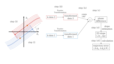 |
A Method for Correcting Non-linear Errors in Radial Trajectories
Hideaki Kutsuna1, Sho Kawajiri2, and Hidenori Takeshima3
1MRI Systems Development Department, Canon Medical Systems Corporation, Kanagawa, Japan, 2MRI Systems Development Department, Canon Medical Systems Corporation, Tochigi, Japan, 3Advanced Technology Research Department, Research and Development Center, Canon Medical Systems Corporation, Kanagawa, Japan The authors propose a new method to use a non-linear model for correcting errors in radial trajectories. The model represents shifts of trajectories as a combination of a linear function and a sigmoid function. Conventional methods assume that the shifts are proportional to the gradient strengths. However, actual trajectories cannot be represented as linear models precisely due to non-linear imperfections of the gradients such as eddy currents. The proposed method suppresses streak artifacts and image shadings which appear with conventional linear correction. |
|||
3525.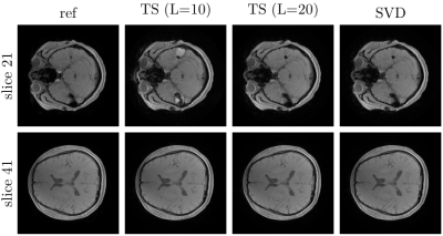 |
Fast Image Reconstruction for Non-Cartesian Acquisitions in the Presence of B0-inhomogeneities
Mirco Grosser1,2 and Tobias Knopp1,2
1Section for Biomedical Imaging, University Medical Center Hamburg-Eppendorf, Hamburg, Germany, 2Institute for Biomedical Imaging, Hamburg University of Technology, Hamburg, Germany
We propose a fast algorithm for the reconstruction of non-cartesian acquisitions with B0-inhomogeneity. The proposed method uses a new SVD-based approximation of the B0-aware imaging operator in combination with diagonal k-space preconditioning. The proposed SVD-based approximation adaptively determines the required number of basis functions and thus reduces the computational effort. Furthermore, we present a method to efficiently compute the $$$\ell_2$$$-optimal diagonal k-space preconditioner taking into account the B0-map. The obtained preconditioner closely matches the one without B0-map. Our experiments demonstrate the fast convergence and reduced computational costs of the proposed method.
|
|||
3526.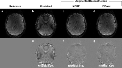 |
FID-navigated phase correction for multi-shot 3D EPI acquisitions
Tess E Wallace1,2, Tobias Kober3,4,5, Simon K Warfield1,2, and Onur Afacan1,2
1Computational Radiology Laboratory, Boston Children's Hospital, Boston, MA, United States, 2Radiology, Harvard Medical School, Boston, MA, United States, 3Advanced Clinical Imaging Technology, Siemens Healthcare, Lausanne, Switzerland, 4Radiology, Lausanne University Hospital and University of Lausanne, Lausanne, Switzerland, 5LTS5, Ecole Polytechnique Federale de Lausanne, Lausanne, Switzerland
Segmented 3D EPI yields higher SNR compared to conventional 2D EPI but is highly sensitive to shot-to-shot B0 variations, which result in image artifacts and compromise the achievable tSNR. In this work, we use FIDnavs combined with an augmented reconstruction strategy to compensate for the adverse effects of spatiotemporal field variations in 3D EPI. We demonstrate that FIDnavs can rapidly and accurately estimate B0 field changes up to second order in three dimensions and that FID-navigated augmented reconstruction results in improved 3D EPI image quality and temporal stability of the BOLD signal time course.
|
|||
3527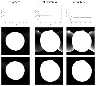 |
Temporal Oscillation in the Phase Error as an Unresolved Source of Ghosting in EPI at 7T Video Permission Withheld
Pål Erik Goa1 and Neil Peter Jerome2
1Dept. of Physics, NTNU, Trondheim, Norway, 2Siemens Healthineers, Trondheim, Norway
Conventional phase-correction algorithms for EPI reconstruction assume constant linear offset in odd-even k-space lines, and so cannot account for dynamically varying offset. The effect of such terms therefore gives rise to Nyquist ghosting. We illustrate this effect with simulated data, and demonstrate the presence of a damped oscillating term in linear phase for ultra-high field (7T) compared to lower field strength (3T). Line-by-line phase correction, using an extended navigator echo train over the full k-space, is able to substantially improve the appearance of ghosting.
|
|||
3528.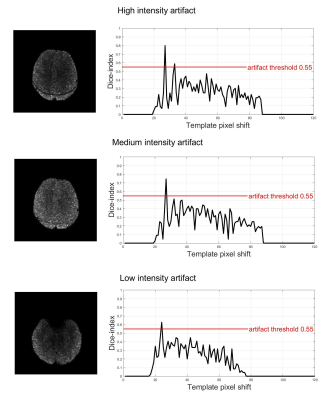 |
Automatic detection of “fat suppression-like” artifacts in brain diffusion MRI
Stefano Tambalo1,2, Riccardo Pederzolli2, Andrea Spagnolo2, Lisa Novello1, and Jorge Jovicich1
1CIMeC, University of Trento, Trento, Italy, 2Department of Radiology, G.B. Rossi Hospital, University of Verona, Verona, Italy
Brain diffusion MRI data may present unpredictable artifacts like those related to insufficient fat suppression. Given the quantity of data collected in state-of-the-art diffusion protocols, such artifacts are challenging to detect by visual inspection. In this study, we implemented and tested an automated method that helps to detect such artifacts, identifying the slice(s) where they are present. The method is based on the automated generation of the shape of the artifact from the skull at each slice and subsequent search of the pattern in the diffusion data. The method gave high specificity (0.977) and sensitivity (0.889).
|
|||
3529.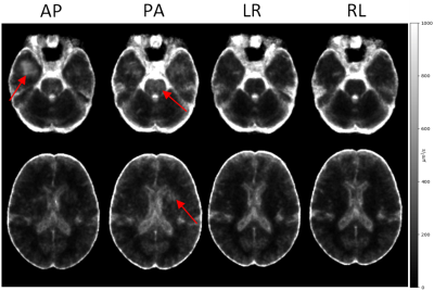 |
Effects of Phase-Encoding Directions on Diffusion MRI Reproducibility
Grayson Clark1, M. Okan Irfanoglu1, and Carlo Pierpaoli1
1QMI, NIBIB/NIH, Bethesda, MD, United States
In this study, we report on the reproducibility of DTI-derived metrics w.r.t. the phase-encoding directions (PED) used for the acquisition of diffusion weighted images. Over the entire brain, the reproducibility of DTI metrics was higher for data acquired using LR/RL phase-encoding directions. However, AP/PA data showed better reproducibility in some regions. The main source of these reproducibility variations was identified as ghosts overlapping with different brain regions depending on PED. We conclude that acquiring data with all four phase-encoding directions would be the most beneficial to achieve maximum reproducibility in all brain regions after proper editing of regionally corrupted data.
|
|||
3530.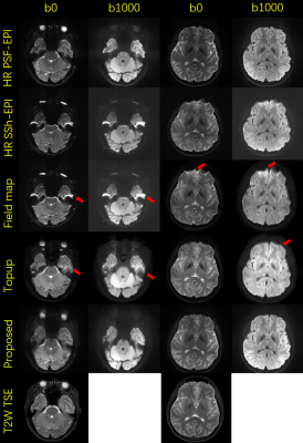 |
Simultaneous super resolution and distortion correction for Single-shot EPI DWI using deep learning method
Xinyu Ye1, Yuan Lian1, Pylypenko Dmytro1, Yajing Zhang2, and Hua Guo1
1Center for Biomedical Imaging Research, Department of Biomedical Engineering, School of Medicine, Tsinghua University, Beijing, China, 2MR Clinical Science, Philips Healthcare, Suzhou, China
Single-shot EPI is widely used for clinical DWI acquisitions. However, due to the limited bandwidth along PE direction, the obtained images suffer from distortion and blurring, which limits its diagnosis capability. Here we propose a deep learning-based method to simultaneously increase resolution and correct distortions for SSh-EPI. In-vivo DWI data are used to test the proposed method. The results show that distortion-corrected high-resolution DWI images can be reconstructed from low-resolution SSh-EPI images and high-resolution anatomical images.
|
|||
3531.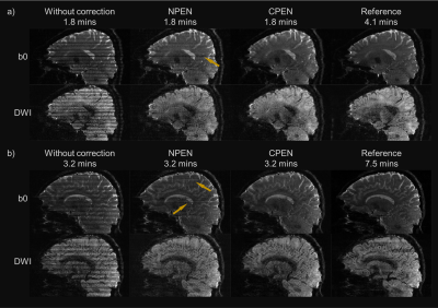 |
Convolutional Neural Network for Slab Profile Encoding (CPEN) in Simultaneous Multi-slab (SMSlab) Diffusion Weighted Imaging
Jieying Zhang1, Simin Liu1, Yuhsuan Wu1, Yajing Zhang2, and Hua Guo1
1Center for Biomedical Imaging Research, Department of Biomedical Engineering, Tsinghua University, Beijing, China, 2MR Clinical Science, Philips Healthcare, Suzhou, China
Simultaneous multi-slab (SMSlab) is a 3D acquisition method that can achieve optimal SNR efficiency for isotropic high-resolution DWI. However, boundary artifacts restrain its application. Nonlinear inversion for slab profile encoding (NPEN) seems to be inadequate for boundary artifacts correction in SMSlab. In this study, we proposed to use a model-based convolutional neural network (referred as CPEN) for this problem. According to the results, it outperforms NPEN in images with different resolutions, and the computation is much faster. Using CPEN, small oversampling factors can be used to reduced the acqsuition time, which is of great meaning for high-resolution whole-brain DWI.
|
|||
3532.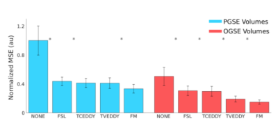 |
Validation of a Retrospective Eddy Current Correction Algorithm for Advanced Diffusion MRI
Paul I Dubovan1,2, Jake J Valsamis1,2, and Corey A Baron1,2
1Medical Biophysics, Western University, London, ON, Canada, 2Center for Functional and Metabolic Mapping, Robarts Research Institute, London, ON, Canada
Diffusion MRI (dMRI) suffers from eddy current-induced distortions which affect the quality of reconstructed images. While existing computational techniques such as FSL eddy can correct most eddy-current induced distortions for traditional techniques, their ability to correct more complex techniques such as Oscillating Gradient Spin Echo (OGSE) remains uncertain due to the time-varying behavior of eddy currents not being considered. We propose an algorithm that considers this behavior and compare its correction quality to other methods. For OGSE acquisitions, the technique outperformed other computational methods that treat eddy currents as static, showing its feasibility as a correction tool for advanced dMRI.
|
|||
3533.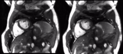 |
Retrospective Eddy Current Artifact Reduction for Balanced SSFP Cine Imaging via Deep Learning
Cynthia Chen1, Christopher Sandino2, Adam Bush3, Frank Ong2, and Shreyas Vasanawala3
1California Institute of Technology, Pasadena, CA, United States, 2Electrical Engineering, Stanford University, Stanford, CA, United States, 3Radiology, Stanford University, Stanford, CA, United States
Eddy currents due to changing magnetic fields reduce diagnostic image quality, especially in SSFP acquisitions. In this work, we propose a deep learning method that successfully reduces eddy current artifacts in 2D cardiac cine imaging using a 3D U-Net architecture. Our method is completely retrospective and does not require any sequence or hardware modifications.
|
|||
3534.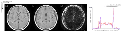 |
Dispersing FID artifact uniformly by modulating phase of 180 degrees pulse of Spin Echo sequence with quadratic function.
Kosuke Ito1 and Atsushi Kuratani1
1Healthcare Business Unit, Hitachi, Ltd., Kashiwa, Japan
T1 weighted image is acquired using Spin Echo sequence in almost clinical routine practices. FID signal induced by 180 degrees pulse causes zipper artifact at the center of the image. To move the artifact to the edge of the image, the phase of RF pulse is controlled as 0 and 180 degrees alternately. However, higher parallel imaging factor cannot be applied due to the FID artifact still exist at the edge of FOV. In this study, FID artifact was dispersed uniformly by modulating phase of 180 degrees pulse with quadratic function, and higher parallel imaging factor was applied in vivo.
|
|||
3535.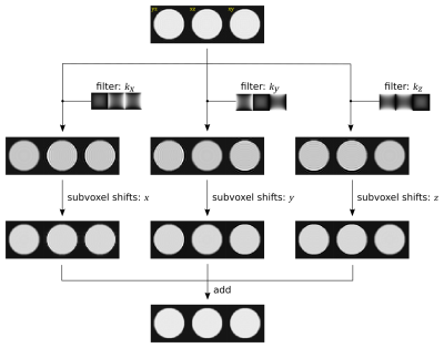 |
Removal of Gibbs ringing artefacts for 3D acquisitions using subvoxel shifts
Thea Bautista1, Jonathan O'Muircheartaigh2,3,4,5, Joseph V Hajnal1,3, and J-Donald Tournier1,3
1Department of Biomedical Engineering, School of Biomedical Engineering and Imaging Sciences, King's College London, London, United Kingdom, 2Forensic & Neurodevelopmental Sciences, King's College London, London, United Kingdom, 3Centre for the Developing Brain, School of Biomedical Engineering and Imaging Sciences, King's College London, London, United Kingdom, 4Department of Perinatal Imaging & Health, School of Biomedical Engineering and Imaging Sciences, King's College London, London, United Kingdom, 5MRC Centre for Neurodevelopmental Dirorders, King's College London, London, United Kingdom
The method of subvoxel shifts is widely used for the removal of Gibbs ringing artefacts from 2D multislice data, but is not appropriate for 3D data. Here, we show that the method can trivially be extended to cater for the 3D case with simple modifications. We demonstrate the method on a numerical phantom, and assess its practical performance using in vivo 3D brain scans.
|
|||
3536.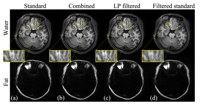 |
Ringing Artifacts Reduction with Low-Pass Filtered Deblurring Kernels
Dinghui Wang1 and James G. Pipe1
1Radiology, Mayo Clinic, Rochester, MN, United States
Spiral imaging has been used in many applications for its fast imaging speed, high scan efficiency and low sensitivity to motion. However, ringing artifacts are often observed in regions where the field map Δf0 changes rapidly in space, especially with long spiral readouts. The goal of this work is to address this issue with a modified deblurring model that weights more on low spatial frequencies. Simulation and volunteered data have demonstrated that the ringing artifacts can be substantially reduced by the proposed method.
|
|||
3537.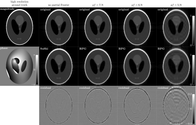 |
Removal of Partial Fourier-Induced Gibbs (RPG) Ringing artifacts in MRI
Hong-Hsi Lee1, Dmitry S Novikov1, and Els Fieremans1
1New York University School of Medicine, New York, NY, United States
Gibbs-ringing artifact in magnitude images obtained by using partial Fourier (PF) acquisition and zero filling interpolation is modeled analytically, and a correction pipeline based on modifying the local subvoxel-shifts method is proposed. With the understanding of oscillating convolution kernel due to the PF acquisition, the ringings in magnitude images can be robustly removed without the need of ad hoc image models and tuning parameters. The effects of ringings on diffusion metrics are further demonstrated in numerical phantoms and in vivo diffusion data. The proposed pipeline removes most ringings in magnitude images and stabilizes estimations of diffusion metrics.
|
The International Society for Magnetic Resonance in Medicine is accredited by the Accreditation Council for Continuing Medical Education to provide continuing medical education for physicians.