Oral
Velocity & Flow
ISMRM & SMRT Annual Meeting • 15-20 May 2021

| Concurrent 4 | 14:00 - 16:00 | Moderators: Petter Dyverfeldt & Eva Peper |
 |
0301.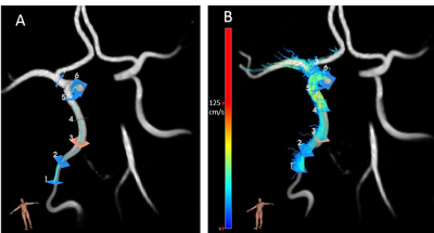 |
Does the internal carotid artery attenuate blood-flow pulsatility in small vessel disease? A 7T 4D-flow MRI study.
Rick J. van Tuijl1, Ynte M. Ruigrok2, Irene C. van der Schaaf1, Lennart J. Geurts1, Gabriël J. E. Rinkel2, Birgitta K. Velthuis1, and Jaco J. M. Zwanenburg1
1Radiology, UMC Utrecht, Utrecht, Netherlands, 2Neurology, UMC Utrecht, Utrecht, Netherlands
We studied blood-flow pulsatility and arterial distensibility along the internal carotid artery (ICA) in cerebral small vessel disease (CSVD) patients and healthy controls using 7Tesla MRI. 4D-flow measurements (0.8 mm isotropic resolution), were analyzed in 17 patients with lacunar infarcts or deep intracerebral hemorrhage (CSVD) and 17 age and sex matched healthy controls. Pulsatility was significantly higher and arterial distensibility significantly lower in CSVD patients compared to controls. Velocity pulsatility was attenuated between the extracranial ICA and the circle of Willis in controls, but increased in CSVD. Higher calcification in CSVD patients correlated with reduced distensibility and increased velocity pulsatility.
|
|
|
0302.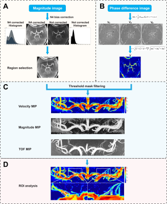 |
Phase-contrast MR angiography at 7 Tesla revealed reduced lenticulostriate artery blood flow velocity in patients with small vessel disease
Yue Wu1,2,3, Chengyue Sun4, Qingle Kong5, Zhixin Li1,2,3, Dongbiao Sun1,2,3, Chen Ling4, Jing An6, Rong Xue1,2,3, Yan Zhuo1,2,3, Yun Yuan4, and Zihao Zhang1,2,3
1State Key Laboratory of Brain and Cognitive Science, Institute of Biophysics, Chinese Academy of Sciences, Beijing, China, 2The Innovation Center of Excellence on Brain Science, Chinese Academy of Sciences, Beijing, China, 3University of Chinese Academy of Sciences, Beijing, China, 4Department of Neurology, Peking University First Hospital, Beijing, China, 5MR Collaboration, Siemens Healthcare Ltd, Beijing, China, 6Siemens Shenzhen Magnetic Resonance Ltd, Shenzhen, China
In this study, we demonstrated a technique that could non-invasively quantify lenticulostriate artery (LSA) flow velocities in cerebral small vessel disease (CSVD). With phase-contrast magnetic resonance angiography (PC-MRA) at 7T, LSA blood flow velocities were detected in patients with CADASIL (a hereditary CSVD). LSA flow velocities decreased in patients compared with healthy individuals. We also found good associations between velocities and clinical characteristics among patients with CADASIL. These results suggest that PC-MRA at 7T is a valuable technique to assess small arterial dysfunction in patients with CSVD.
|
|
0303.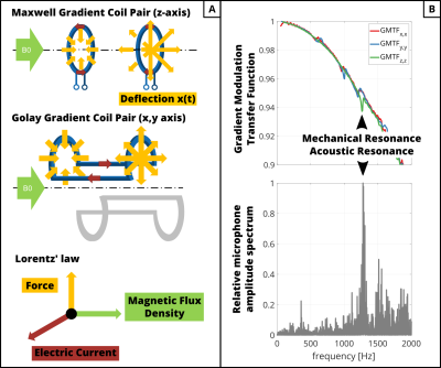 |
Background Phase Error Reduction in Phase-Contrast MRI based on Acoustic Noise Recordings
Hannes Dillinger1, Eva Peper1, Christian Guenthner1, and Sebastian Kozerke1
1Institute for Biomedical Engineering, University and ETH Zurich, Zurich, Switzerland
This work identifies mechanical resonances of the gradient coil system as a source of increased spatially linear and quadratic phase offsets. Mechanical resonance frequencies were identified by mobile phone audio recordings from inside the scanner room and compared to the gradient modulation transfer function. Results demonstrate that optimal TE reduces phase ramps by a factor of 15 and optimal TR removes the spatially quadratic phase offset.
|
||
|
0304.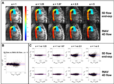 |
Correcting vs resolving respiratory motion in accelerated free-running whole-heart radial flow MRI using focused navigation (fNAV)
Mariana Baginha da Lança Falcão1, Giulia M. C. Rossi1, Liliana Ma2,3, John Heerfordt1,4, Davide Piccini1,4, Jérôme Yerly1,5, Milan Prša6, Tobias Rutz7, Estelle Tenisch1, Michael Markl2,3, Matthias Stuber1,5, and Christopher W. Roy1
1Department of Radiology, Lausanne University Hospital (CHUV) and University of Lausanne (UNIL), Lausanne, Switzerland, 2Department of Radiology, Feinberg School of Medicine, Northwestern University, Chicago, IL, United States, 3Department of Biomedical Engineering, Northwestern University, Chicago, IL, United States, 4Advanced clinical imaging technology, Siemens Healthcare AG, Lausanne, Switzerland, 5Center for Biomedical Imaging (CIBM), Lausanne, Switzerland, 6Woman-Mother-Child Department, Lausanne University Hospital (CHUV) and University of Lausanne (UNIL), Lausanne, Switzerland, 7Service of Cardiology, Centre de resonance magnétique cardiaque (CRMC), Lausanne University Hospital (CHUV) and University of Lausanne (UNIL), Lausanne, Switzerland
In this work, a free-running radial whole-heart flow sequence was acquired in five congenital heart disease patients and images were reconstructed using a) a previously developed 5D flow framework for respiratory and cardiac resolved images, and b) a novel framework for respiratory motion corrected and cardiac resolved 4D flow (fNAV). Image and flow differences were measured across a range of acceleration factors. We showed that the free-running acquisition, which is already undersampled, can be even further accelerated with less signal degradation if it is reconstructed with fNAV 4D flow, compared to using 5D flow.
|
|
0305.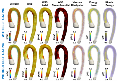 |
Impact of respiratory Gating on hemodynamic parameters from 4D flow MRI
Esteban Jorge Denecken-Campaña1,2,3, Julio Sotelo1,3,4, Cristobal Arrieta1,3, Pablo Irarrazaval1,2,3, Cristián Tejos1,2,3, Marcelo E. Andia1,3,5, and Sergio Uribe1,3,5
1Biomedical Imaging Center, Pontificia Universidad Católica de Chile, Santiago, Chile, 2Electrical Engineering Department, School of Engineering, Pontificia Universidad Católica de Chile, Santiago, Chile, 3ANID – Millennium Science Initiative Program – Millennium Nucleus for Cardiovascular Magnetic Resonance, Santiago, Chile, 4School of Biomedical Engineering, Universidad de Valparaíso, Valparaíso, Chile, 5Department of Radiology, School of Medicine, Pontificia Universidad Católica de Chile, Santiago, Chile
Hemodynamic parameters from 4D flow datasets derived in the last years have shown promising diagnostic value in different cardiovascular pathologies. However, we know little about the behavior of these parameters when the 4D flow data is corrupted by respiratory motion. The purpose of this work is to perform a quantitative comparison between hemodynamic parameters computed from 4D flow Cardiac MRI with and without respiratory self-gating. We found significant variability of the hemodynamic parameters in the ascending aorta of healthy volunteers when comparing both methods. Hemodynamic parameters measured with self-gating acquisition showed statistically significant differences compared to those measured without self-gating.
|
||
|
0306.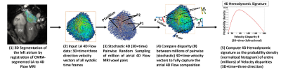 |
Novel Stochastic 4D Flow Signatures of time-resolved 3D left atrial flow-field alterations in atrial fibrillation
Thara Nallamothu1,2, Amanda L. DiCarlo1, Daniel C. Lee3, Daniel Kim1, Rishi Arora3, Michael Markl1,2, Phillip Greenland4, Rod Passman3, and Mohammed S.M. Elbaz1
1Radiology, Northwestern University, Chicago, IL, United States, 2Biomedical Engineering, Northwestern University, Chicago, IL, United States, 3Medicine (Cardiology), Northwestern University, Chicago, IL, United States, 4Preventative Medicine, Northwestern University, Chicago, IL, United States
4D Flow MRI studies have shown an association between atrial fibrillation (AF) with altered left atrial (LA) blood flow. Nevertheless, LA flow dynamics changes can be complex (vortex flow, jets, stagnations, etc.). Existing quantitative 4D flow metrics only characterize parts of the overall complex interacting LA flow dynamics. Here, we propose a novel stochastic 4D Flow signature technique to quantify the unique composition of normal and altered LA flow dynamics utilizing the entire 4D three-directional velocity-field from 4D Flow MRI. We demonstrate the excellent reproducibility and the feasibility of the technique in quantifying distinctly altered LA signatures in AF patients.
|
|
0307.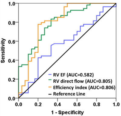 |
Insight of right ventricular dysfunction and impaired efficiency via 4D flow CMR in repaired tetralogy of Fallot
Xiaodan Zhao1, Liwei Hu2, Ru-San Tan1,3, Ping Chai4, Marielle Fortier3,5, Rong Zhen Ouyang2, Shuo Zhang6, Wen Ruan1, Ting Ting Low4, Shuang Leng1, Jun-Mei Zhang1,3, Bryant Jennifer1, Lynette Teo4, Rob van der Geest7,
Teng Hong Tan3,5, James W. Yip4, Ju Le Tan1,3, Yumin Zhong2, and Liang Zhong1,3
1National Heart Centre Singapore, Singapore, Singapore, 2Shanghai Children’s Medical Centre, Shanghai, China, 3Duke-NUS Medical School, Singapore, Singapore, 4National University Hospital Singapore, Singapore, Singapore, 5KK Women’s and Children’s Hospital, Singapore, Singapore, 6Philips Germany, Humburg, Germany, 7Leiden University Medical Center, Leiden, Netherlands
4D flow CMR enables qualitative and quantitative assessment of intra-cardiac flow. Kinetic energy (KE) and pathline-derived four flow components for left ventricular (LV) and right ventricular (RV) were analyzed and compared in repaired tetralogy of Fallot (rTOF) and age-matched controls. For RV, rTOF had increased peak systolic, systolic and peak E-wave KE normalized to end-diastolic volume while decreased efficiency index. RV direct flow decreased while RV residual volume increased from controls to rTOF with preserved RVEF (rTOFpEF) to rTOF with reduced RVEF. ROC analysis showed RV direct flow and efficiency index were sensitive markers to detect RV dysfunction in rTOFpEF.
|
||
 |
0308.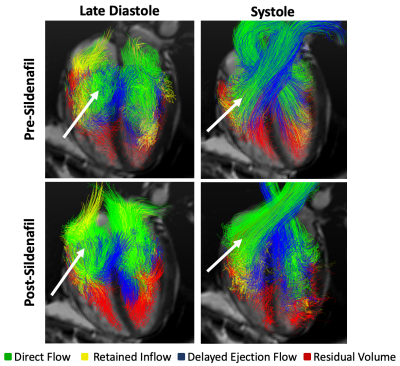 |
Sildenafil Administration Improves Right Ventricular Function on 4D Flow MRI in Young Adults Born Premature
Philip A Corrado1, Gregory P Barton2, Christopher J François3, Oliver Wieben1, and Kara N Goss2
1University of Wisconsin-Madison, Madison, WI, United States, 2University of Texas Southwestern, Dallas, TX, United States, 3Mayo Clinic, Rochester, MN, United States
We used 4D flow MRI before and during acute pharmacological intervention to reduce either afterload or heart rate in young adults born very to extremely premature, finding improved overall cardiac function and shifted intraventricular flow in the RV towards direct flow after RV afterload reduction with sildenafil. We interpret these findings to mean that intrinsic morphologic differences as well as increased RV afterload are stronger drivers of cardiac dysfunction in the preterm heart than decreased filling time. This study design may serve as a blueprint for future studies investigating the effects of acute hemodynamic pharmacological interventions with 4D flow MRI.
|
|
|
0309.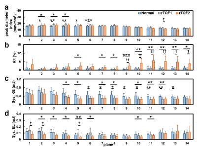 |
Abnormal aortic kinetic energy and viscous energy loss in patients with repaired tetralogy of Fallot
Yu-Ru Yang1, Meng-Chu Chang1, Ming-Ting Wu2, Ken-Pen Weng3, and Hsu-Hsia Peng1
1Biomedical Engineering and Environmental Sciences, National Tsing Hua University, Hsinchu, Taiwan, 2Radiology, Kaohsiung Veterans General Hospital, Kaohsiung, Taiwan, 3Pediatrics, Kaohsiung Veterans General Hospital, Kaohsiung, Taiwan
We aim to evaluate aortic kinetic energy (KE) and viscous energy loss (EL) in repaired tetralogy of Fallot (rTOF) patients with different degrees of aortic regurgitation fraction (RF). The rTOF1 group (RF<2%) demonstrated decreased systolic KE in arch and descending aorta, suggesting the mild altered aortic flow. The rTOF2 group (RF≧2%) presented significantly elevated RF in arch and descending aorta, increased systolic KE from distal ascending aorta to proximal descending aorta, and decreased systolic EL in ascending aorta and distal arch. In conclusion, the systolic KE may provide earlier evidence of abnormal aortic flow before serious aortic regurgitation.
|
|
0310.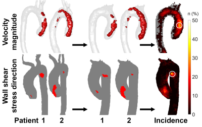 |
Abnormal aortic hemodynamics at predilection sites for dissection in Marfan patients: a 4D flow study
Pim van Ooij1, Mitzi van Andel2, Lukas M. Gottwald1, Aart J Nederveen1, and Maarten Groenink1
1Radiology & Nuclear Medicine, Amsterdam University Medical Centers, location AMC, Amsterdam, Netherlands, 2Cardiology, Amsterdam University Medical Centers, location AMC, Amsterdam, Netherlands
In this study we use 4D flow MRI techniques to investigate abnormal magnitude and direction of blood flow velocity and wall shear stress in patients with Marfan syndrome, a congenital disease that may cause aortic dissection. We found that patients that underwent aortic root repair have significantly more abnormal hemodynamics and that abnormally elevated hemodynamics were associated with blood pressure chracteristics. Abnormally directed hemodynamics were not associated with any patient characteristics, but showed a distinct regional increase at the inner proximal descending aorta, awell-know predilection site for aortic dissection in Marfan patients.
|
The International Society for Magnetic Resonance in Medicine is accredited by the Accreditation Council for Continuing Medical Education to provide continuing medical education for physicians.