Oral
Artifacts & Corrections
ISMRM & SMRT Annual Meeting • 15-20 May 2021

| Concurrent 1 | 12:00 - 14:00 | Moderators: Christopher Wiggins |
0665.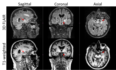 |
Left-Right Intensity Asymmetries Systematically Vary Across MR Scanners and Introduce Diagnostic Uncertainty
Arvin Arani1, Christopher G. Schwarz1, Matthew C. Murphy1, Joshua D. Trzasko1, Jeffrey L. Gunter1, Matthew L. Senjem1, Heather J. Wiste1, Kiaran P. McGee1, Matthew A. Bernstein1, John Huston III1, and Clifford R. Jack Jr.1
1Mayo Clinic, Rochester, MN, United States
In magnetic resonance imaging (MRI) many factors can contribute to non-tissue specific image intensity inhomogeneity. However, the potential clinical impact or systematic biases of these effects have not been extensively investigated across multiple MRI vendors and models for neuroimaging applications. Specifically, left-right intensity comparisons are commonly used by radiologists to verify/identify pathology. If significant systematic left-right intensity asymmetries (LRIA) exist, it may lead to diagnostic uncertainty and result in unnecessary imaging follow-up and patient burden. This study shows that LRIA are common, system specific, systematic, can mimic disease, create diagnostic uncertainty, and can impact multiple sequences (T1-weighted and FLAIR).
|
||
 |
0666.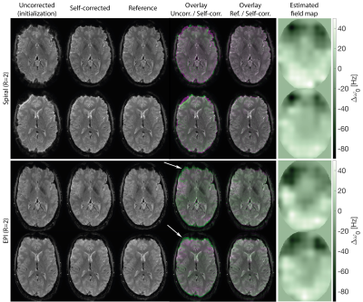 |
Off-Resonance Self-Correction by Implicit B0-Encoding
Franz Patzig1, Bertram Wilm1, and Klaas Paul Pruessmann1
1Institut for Biomedical Engineering, ETH Zurich and University of Zurich, Zurich, Switzerland
A caveat in using trajectories with long readout durations are artefacts due to B0 inhomogeneity. Correcting these based on B0-maps or reverse-phase-encoded EPI requires additional scan time and is unattractive for some applications. In this work, the capability of coil-arrays to extrapolate information in k-space is shown to also allow the extraction of temporal information such as the time-varying phase introduced by B0-offsets. An optimization problem is formulated to retrieve an estimate of B0 from measured data without making assumptions on the employed trajectory. B0 estimation and self-correction of single-shot spiral and EPI in-vivo data (up to R=4) is demonstrated.
|
|
0667. |
Improved Fat and Water Depiction in Musculoskeletal MRI by Control of Through-Slice Chemical-Shift Artifacts in 2D Turbo-Spin-Echo Imaging at 7 T
Constantin von Deuster1,2, Stefan Sommer1,2, Christoph Germann3,4, Natalie Hinterholzer2, Robin M. Heidemann5, Reto Sutter3,4, and Daniel Nanz2,4
1Siemens Healthcare, Zurich, Switzerland, 2Swiss Center for Musculoskeletal Imaging (SCMI), Balgrist Campus, Zurich, Switzerland, 3Radiology, Balgrist University Hospital, Zurich, Switzerland, 4University of Zurich, Zurich, Switzerland, 5Siemens Healthcare, Erlangen, Germany
The large water-fat frequency difference at 7 T renders imaging of the musculoskeletal (MSK) anatomy very challenging. In particular, through-slice chemical-shift artifacts may manifest in state-of-the-art 2D turbo-spin-echo (TSE) images as partial or locally complete fat-signal loss that radiologists are usually not trained to account for from lower field strengths. In this work, we demonstrate the range of possible through-slice artifacts in MSK images and show that matched RF-pulse bandwidths as high as 1500 Hz for the excitation and refocusing RF-pulses are necessary to consistently perform successful, non-fat suppressed MSK imaging at 7 T.
|
||
 |
0668. |
Concomitant field compensation using additional oscillating gradients in a double diffusion encoding imaging sequence
Julian Rauch1,2, Frederik B. Laun3, Theresa Palm3, Jan Martin3,4, Maxim Zaitsev5,6, Mark E. Ladd1,2,7, Peter Bachert1,2, and Tristan A. Kuder1
1Division of Medical Physics in Radiology, German Cancer Research Center (DKFZ), Heidelberg, Germany, 2Faculty of Physics and Astronomy, Heidelberg University, Heidelberg, Germany, 3Institute of Radiology, University Hospital Erlangen, Friedrich-Alexander-Universität Erlangen-Nürnberg (FAU), Erlangen, Germany, 4Division of Physical Chemistry, Lund University, Lund, Sweden, 5Medical Physics, Department of Radiology, Faculty of Medicine, Medical Center University of Freiburg, Freiburg, Germany, 6High Field Magnetic Resonance Center, Center for Medical Physics and Biomedical Engineering Medical University of Vienna, Vienna, Austria, 7Faculty of Medicine, Heidelberg University, Heidelberg, Germany
Concomitant or Maxwell fields cause intravoxel dephasing which can lead to strong image artifacts. In this study, we present a new method for concomitant field correction in double diffusion encoding sequences with single pairs of bipolar gradients on each axis. Additionally implemented oscillating gradients remove the dephasing without changing the desired image. Phase and magnitude images are analyzed with respect to concomitant field induced artifacts and the proposed correction method. We show that the compensation eliminates these artifacts without further consequences for image quality. The method also may be included in other imaging sequences to achieve concomitant field compensation.
|
|
0669.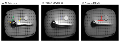 |
Spectrally-encoded multi-spectral imaging (SEMSI) for off-resonance correction near metallic implants.
Daehyun Yoon1, Philip Lee2, Krishna Nayak3, and Brian Hargreaves1
1Radiology, Stanford University, Stanford, CA, United States, 2Electrical Engineering, Stanford University, Stanford, United Kingdom, 3Electrical and Computer Engineering, University of Southern California, Los Angeles, CA, United States
Multi-spectral imaging (MSI) including view-angle-tilting (VAT) is the dominant technique to correct for severe off-resonance artifacts near metallic implants. While VAT mitigates the signal pile-up and translation artifact in the area of severe off-resonance, it also causes global blurring and SNR loss in the on-resonance area away from metal. In this work, we introduce a novel spectrally encoded MSI approach, denoted SEMSI, that resolves pile-up and translation artifacts without VAT or z-phase encoding. Phantom imaging results show the promise of SEMSI to provide high-quality, artifact-free images in the presence of metallic implants without global blurring.
|
||
|
0670.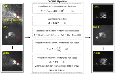 |
Cancellation of streak artifacts using the interference null space (CACTUS) for radial abdominal imaging
Zhiyang Fu1,2, Maria I Altbach1,3, and Ali Bilgin1,2,3
1Department of Medical Imaging, University of Arizona, Tucson, AZ, United States, 2Department of Electrical and Computer Engineering, University of Arizona, Tucson, AZ, United States, 3Department of Biomedical Engineering, University of Arizona, Tucson, AZ, United States
In radial imaging, streaks due to gradient inhomogeneities can appear even when Nyquist criterion is fulfilled. Earlier techniques targeting this type of streaks remove coils contributing prominent streaks at the cost of signal loss. A phased array beamforming based technique (B-STAR), that can achieve a good trade-off between streak reduction and signal preservation, was proposed as a post-processing method. We propose CACTUS, an alternative technique for streak cancellation, which can be used either as a preprocessing method or with iterative reconstructions. In vivo abdominal experiments show enhanced image reconstructions and improved quantitative parameter maps.
|
|
0671.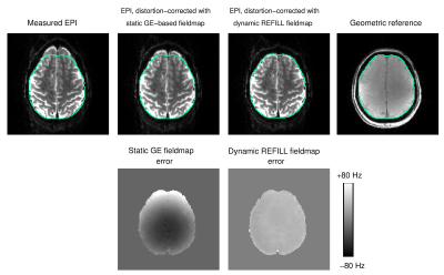 |
Improved dynamic distortion correction for fMRI using single-echo EPI, a fast sensitivity scan and readout-reversed first image (REFILL)
Simon Daniel Robinson1,2,3, Beata Bachrata3,4, Korbinian Eckstein3, Saskia Bollmann1, Steffen Bollmann5, Siegfried Trattnig3, Christian Enzinger2, and Markus Barth5
1Centre for Advanced Imaging, University of Queensland, Brisbane, Australia, 2Department of Neurology, Medical University of Graz, Graz, Austria, 3Department of Biomedical Imaging and Image-guided Therapy, Medical University of Vienna, Vienna, Austria, 4Christian Doppler Laboratory for Clinical Molecular MR Imaging, Medical University of Vienna, Vienna, Austria, 5School of Electrical Engineering and Information Technology, University of Queensland, Brisbane, Australia
We propose an improved dynamic distortion correction method for fMRI. A fast (4s) multi-echo GE reference scan is used to calculate coil sensitivities and other non-ΔB0-related contributions to coil phase, and one EPI volume in which the readout direction is reversed allows a phase gradient in the readout direction which is specific to EPI to be determined. Knowledge of these quantities allow fieldmaps to be calculated from the phase of each single-echo EPI volume. Reverse-Encoded First Image and Low resoLution reference scan (REFILL) fieldmaps accurately measure ΔB0 as it changes due to motion and respiration.
|
||
|
0672.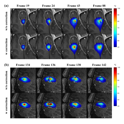 |
Laser Heating Induced Susceptibility Artifacts Cause Significant Temperature Erros in PRF Shift-based MR Thermometry
Ziyi Pan1, Meng Han2, Yawei Kuang2, Hao Sun2, Kai Zhang3, Yuan Lian1, Yishi Wang4, Wenbo Liu2, Guangzhi Wang5, and Hua Guo1
1Center for Biomedical Imaging Research, Department of Biomedical Engineering, School of Medicine, Tsinghua University, Beijing, China, 2Sinovation Medical, Beijing, China, 3Beijing Tiantan Hospital, Capital Medical University, Beijing, China, 4Philips Healthcare, Beijing, China, 5Department of Biomedical Engineering, School of Medicine, Tsinghua University, Beijing, China
MR-guided laser-interstitial thermal therapy (MRgLITT) is a minimally invasive therapeutic method that has created new options for surgically challenging lesions. Most MRgLITT procedures depend on proton-resonance-frequency (PRF) shift-based MR thermometry. However, it can be hampered by magnetic susceptibility changes generated during laser ablation. In this work, we demonstrate for the first time that laser-heating induced susceptibility changes can lead to significant temperature errors, with ex-vivo (pig muscle and brain tissues), in-vivo (Doberman) and clinical (epilepsy patient) experiments. A new algorithm based on multi-echo GRE instead of the conventional single-echo GRE is also introduce to correct the susceptibility-induced temperature errors.
|
|
|
0673.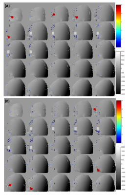 |
Identifying the source of spurious echoes in single voxel 1H MR Spectroscopy
Zahra Shams1, Dennis W.J. Klomp1, Vincent O. Boer2, Jannie P. Wijnen1, and Evita C. Wiegers1
1Department of radiology, University Medical Center Utrecht, Utrecht, Netherlands, 2Danish Research Centre for Magnetic Resonance, Centre for Functional and Diagnostic Imaging and Research, Copenhagen University Hospital Hvidovre, Hvidovre, Denmark
In this work, we proposed a new strategy to identify the source of potential artifacts in single voxel 1H MRS by taking into account the intrinsic B0 field gradients in the human brain. We mapped the intrinsic gradient fields inside the human head to assess the level of signal crushing of each pathway in the entire field of view of the receiver coil. This will enable subject and location specific design of optimal crusher gradient scheme in SV MRS.
|
|
0674.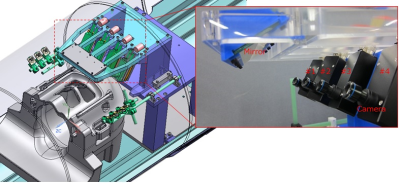 |
Markerless optical head tracking system using facial features
Toru Sasaki1, Ryuichi Nanaumi1, Mitsuo Nishimura1, Kazuhiko Fukutani1, Shuichi Kobayashi1, Kazuya Okamoto2, Hiroshi Kusahara2, and Kazuto Nakabayashi2
1Medical Products Technology Development Center, R&D Headquarters, Canon Inc., Tokyo, Japan, 2Advanced MRI Development PJ Team, Canon Medical Systems Corp., Kanagawa, Japan
We have developed a markerless tracking system enabling to estimate head pose through limited apertures of a head coil. The system, composed of a mirror and four MR compatible cameras, tracks the patient's facial features. The patient’s face image through the head coil’s apertures is monitored during scanning. To demonstrate its feasibility for tracking, a volunteer was moving his head arbitrarily inside a scanner. Despite the limited apertures of the head coil, the facial features were successfully tracked.
|
The International Society for Magnetic Resonance in Medicine is accredited by the Accreditation Council for Continuing Medical Education to provide continuing medical education for physicians.