SMRT Edu Session
MR in Oncology II
ISMRM & SMRT Annual Meeting • 15-20 May 2021

| SMRT Session | 06:00 - 08:00 | Moderators: Jacob Cameron |
 |
Tumour Heterogeneity in Ovarian & Kidney Cancers
Evis Sala
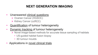
Intra-tumour heterogeneity is present on various levels in ovarian and kidney cancers. Different tumour regions are genetically heterogeneous, show a variable physiology, different metabolic profiles and different morphological appearances. Each of these properties can be investigated in isolation, done typically in the past. Our aim is, however, to understand where Imaging, Transcriptomics, Genomics and Metabolomics provide complementary information and where information is redundant. For this, we require spatially resolved data integration which is key to biological understanding of what we see on imaging as well as for non-invasive evaluation of tumour micro-environment and better prediction of treatment response and outcome.
|
|
| Diffusion & DCE in Chemotherapy Response
Sungheon Gene Kim
Diffusion and DCE-MRI have become important techniques in various areas of cancer imaging including diagnosis, tumor grading, and treatment response evaluation and prediction. The rapid development of new diffusion and perfusion techniques owing to the recent advance in MR hardware and emerging new microstructure models have shown a promising trend to expand the scope of dMRI and DCE-MRI to become a powerful tool in cancer imaging to study tumor heterogeneity, vascularity, cellularity, and microstructural properties. Diffusion and DCE-MRI can provide quantitative measurement of antiangiogenic and cytotoxic effect of chemotherapy.
|
||
| MR-Guided Radiotherapy
Glenn Cahoon
Radiotherapy is one of the most effective cancer treatments available with almost half of all cancer patients receive radiation treatment over the course of their disease. With an increasing number of people surviving cancer, emphasis is being placed on reducing treatment side effects. Improvements in radiation treatment planning and delivery are reliant on accurately visualizing and localising the disease as well as normal tissue structures. Traditionally CT imaging has been used for treatment planning and dose calculation, however, MRI with its superior soft tissue contrast, is increasingly being incorporated into the RT workflow to improve lesion definition and disease extent. |
The International Society for Magnetic Resonance in Medicine is accredited by the Accreditation Council for Continuing Medical Education to provide continuing medical education for physicians.