Yao-Chia Shih1,2,3, Fa-Hsuan Lin4,5, Aeden Kuek Zi Cheng1, Horng-Huei Liou6,7, and Wen-Yih Isaac Tseng3,7,8,9
1Department of Diagnostic Radiology, Singapore General Hospital, Singapore, Singapore, 2Duke-NUS Medical School, Singapore, Singapore, 3Institute of Medical Device and Imaging, College of Medicine, National Taiwan University, Taipei, Taiwan, 4Department of Medical Biophysics, University of Toronto, Toronto, ON, Canada, 5Physical Sciences Platform, Sunnybrook Research Institute, Toronto, ON, Canada, 6Department of Neurology, National Taiwan University Hospital and College of Medicine, Taipei, Taiwan, 7Graduate Institute of Brain and Mind Sciences, College of Medicine, National Taiwan University, Taipei, Taiwan, 8Department of Medical Imaging, National Taiwan University Hospital and College of Medicine, Taipei, Taiwan, 9Molecular Imaging Center, National Taiwan University, Taipei, Taiwan
1Department of Diagnostic Radiology, Singapore General Hospital, Singapore, Singapore, 2Duke-NUS Medical School, Singapore, Singapore, 3Institute of Medical Device and Imaging, College of Medicine, National Taiwan University, Taipei, Taiwan, 4Department of Medical Biophysics, University of Toronto, Toronto, ON, Canada, 5Physical Sciences Platform, Sunnybrook Research Institute, Toronto, ON, Canada, 6Department of Neurology, National Taiwan University Hospital and College of Medicine, Taipei, Taiwan, 7Graduate Institute of Brain and Mind Sciences, College of Medicine, National Taiwan University, Taipei, Taiwan, 8Department of Medical Imaging, National Taiwan University Hospital and College of Medicine, Taipei, Taiwan, 9Molecular Imaging Center, National Taiwan University, Taipei, Taiwan
We identified functional alterations on the paths connecting to the
mammillary body in the simplified hippocampal–diencephalic–cingulate model that
associated with seizure frequency in patients with left and right mesial
temporal sclerosis.

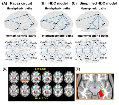
Fig. 1. (A-C) The three candidate neural models.
(D) Regions of interest (ROIs) of the three models were obtained from the
Talairach Daemon atlas in the Montreal Neurological Institute space. (E) The
brown window indicates the anatomical locations of the left hippocampus (HP, green
ROI) and mammillary body (MB, purple ROI). Cyan ROI: anterior cingulate gyrus
(ACG), blue ROI: anterior thalamic nuclei (ATN), red ROI: parahippocampal gyrus
(PHG), yellow ROI: posterior cingulate gyrus (PCG).
