Nicolò Rolandi1, Fulvia Palesi1,2, Francesco Padelli3, Isabella Giachetti3, Domenico Acquino3, Paul Summers4, Giancarlo Germani4, Gerardo Salvato1,5,6, Valeria Mariani5, Pina Scarpa5,6, Egidio D'Angelo1,2, Gabriella Bottini1,5,6, Laura Tassi5, Paolo Vitali4,7, and Claudia Angela Michela Gandini Wheeler-Kingshott1,2,8
1Department of Brain and Behavioral Science, University of Pavia, Pavia, Italy, 2Brain Connectivity Center Research Deparment, IRCCS Mondino Foundation, Pavia, Italy, 3I.R.C.C.S. Istituto Neurologico Carlo Besta, Milano, Italy, 4Neuroradiology Unit, IRCCS Mondino Foundation, Pavia, Italy, 5Hospital Niguarda, Milano, Italy, 6Milan Center for Neuroscience, Milano, Italy, 7Department of Radiology, IRCCS Policlinico San Donato, Milano, Italy, 8NMR Research Unit, Queen Square MS Centre, Department of Neuroinflammation, UCL Queen Square Institute of Neurology, Faculty of brain Sciences, University College London (UCL), London, United Kingdom
1Department of Brain and Behavioral Science, University of Pavia, Pavia, Italy, 2Brain Connectivity Center Research Deparment, IRCCS Mondino Foundation, Pavia, Italy, 3I.R.C.C.S. Istituto Neurologico Carlo Besta, Milano, Italy, 4Neuroradiology Unit, IRCCS Mondino Foundation, Pavia, Italy, 5Hospital Niguarda, Milano, Italy, 6Milan Center for Neuroscience, Milano, Italy, 7Department of Radiology, IRCCS Policlinico San Donato, Milano, Italy, 8NMR Research Unit, Queen Square MS Centre, Department of Neuroinflammation, UCL Queen Square Institute of Neurology, Faculty of brain Sciences, University College London (UCL), London, United Kingdom
Righ TLE showed more alterations than LeftTLE, this finding is in contrast with previous work. Uncinate fasciculus seems to be the most affected tract. ODI in Cingulum showed a negative correlation with both neuropsychological scores.

Figure1: From left to right, axial sagittal coronal view of tracts reconstruction of cingulum (blue), superior longitudinal fasciculus (purple), inferior longitudinal fasciculus (yellow), uncinate fasciculus (light blue).
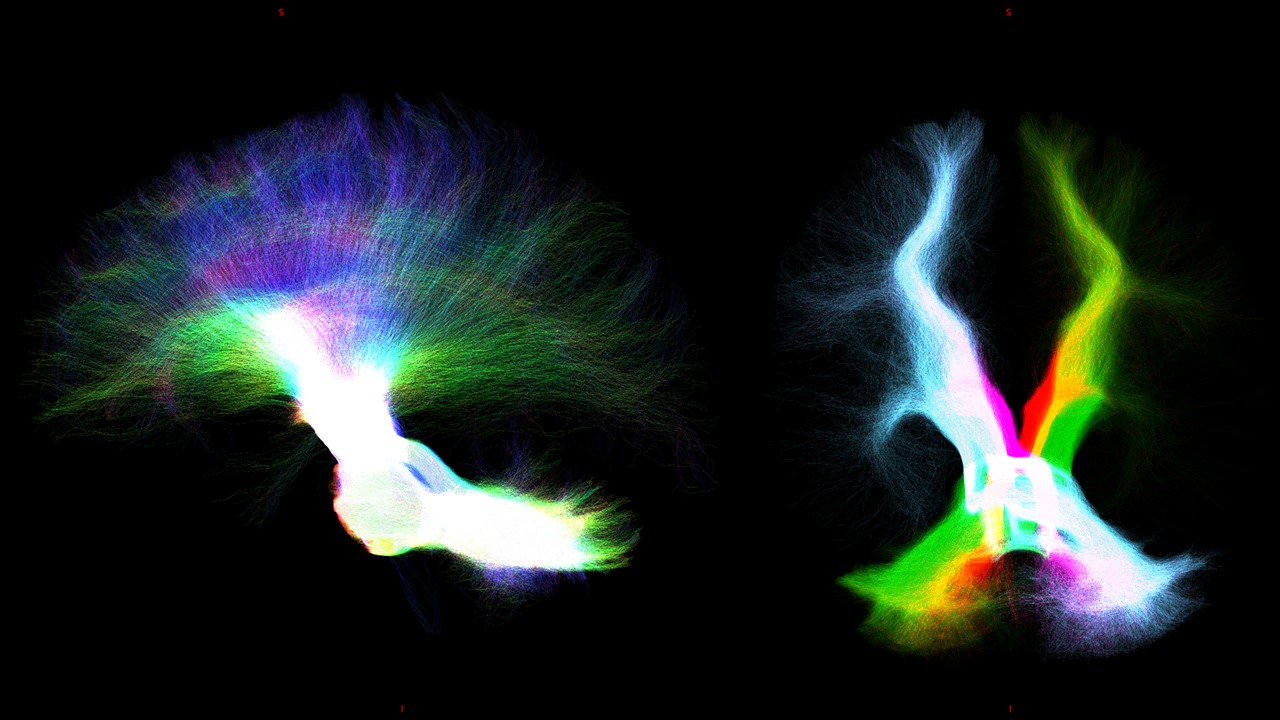
Figure 2: From left to right, sagittal (rgb color direction) and coronal view of tracts reconstruction of cerebello-thalamo-corticalmand (left purple and rightt red) cerebro-ponto-cerebellar tract (left light blue and right green).