Seung-Yi Lee1, Briana Meyer1, Shekar Kurpad2, and Matthew Budde2
1Biophysics, Medical College of Wisconsin, Milwaukee, WI, United States, 2Neurosurgery, Medical College of Wisconsin, Milwaukee, WI, United States
1Biophysics, Medical College of Wisconsin, Milwaukee, WI, United States, 2Neurosurgery, Medical College of Wisconsin, Milwaukee, WI, United States
We show feasibility of high-quality sagittal plane diffusion imaging when
combined with a higher order motion compensation diffusion preparation, both respiratory,
cardiac gating and 2-dimensional filtered diffusion weighted imaging scheme.
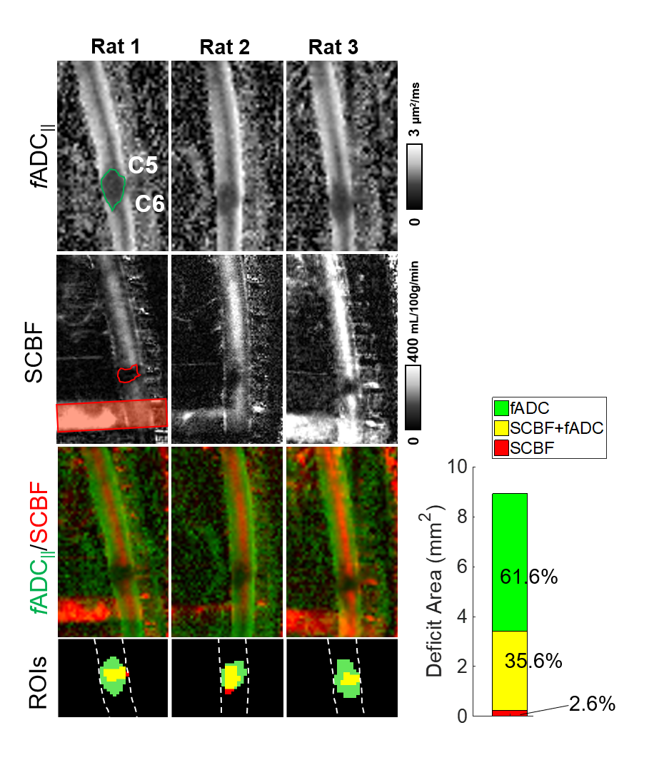
Figure 5 Mismatch of perfusion and diffusion after acute spinal
cord injury. Maps of filtered axial diffusivity (fADC||) and spinal cord blood flow (SCBF) are shown for
three acutely injured animals with the labeling plane (red) indicated. Areas of
decreased fADC|| (green) or SCBF (red) were manually outlined
and along with maps of lesion overlap mean lesion values reflect the extent of
each contrast or overlap. Across three
animals, the perfusion abnormality was clearly and consistently smaller than the
diffusion abnormality.
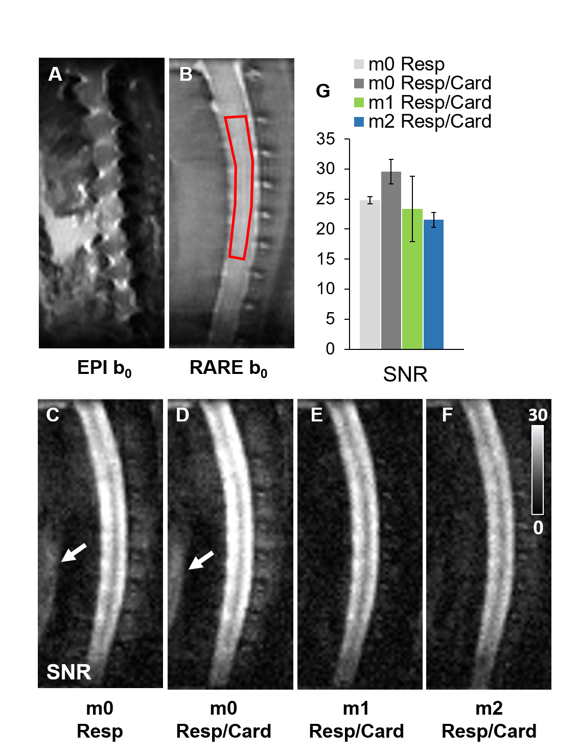
Figure 2 Image quality of sagittal DWprep-RARE in
the spinal cord. Prominent artifacts exist in the EPI image (A) without
diffusion weighting (b0) that are not apparent in the RARE (B),
noting these are from different animals. With b(perpendicular) =2000 s/mm2, subtle artifacts were
evident in both respiratory (C) and dual (D) gating conditions with both m1 (E)
and m2 (F) compensation eliminating the artifacts. Data shown for n=4 animals
obtained from ROIs within the spinal cord as shown (B).