Yiran Li1, Danfeng Xie1, Dushyant Kumar2, Abigail Cember2, Ravi Prakash Reddy Nanga2, Hari Hariharan2, John A. Detre3, Ravinder Reddy2, and Ze Wang1
1Department of Diagnostic Radiology and Nuclear Medicine, University of Maryland School of Medicine, Baltimore, MD, United States, 2Department of Radiology, University of Pennsylvania Perelman School of Medicine, Philadelphia, PA, United States, 3Department of Neurology, University of Pennsylvania Perelman School of Medicine, Philadelphia, PA, United States
1Department of Diagnostic Radiology and Nuclear Medicine, University of Maryland School of Medicine, Baltimore, MD, United States, 2Department of Radiology, University of Pennsylvania Perelman School of Medicine, Philadelphia, PA, United States, 3Department of Neurology, University of Pennsylvania Perelman School of Medicine, Philadelphia, PA, United States
This study presents a DL based framework for correcting B0 inhomogeneity for GluCEST imaging using fewer acquisitions. Based on 3 or 5 positive offset CEST images, the proposed method can save >80% of CEST imaging acquisition time as compared to the conventional protocol.
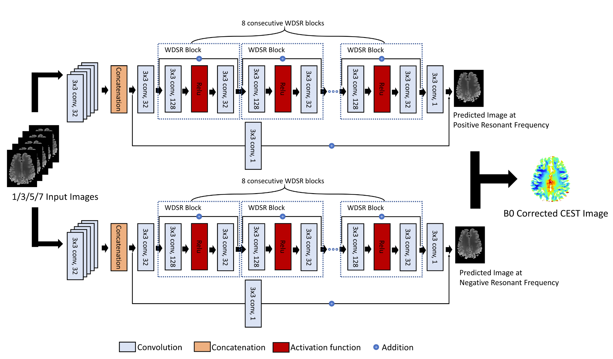
Fig. 2 The
architecture of DL-B0GluCEST-HS is an enhanced deep residual
network. The first layer consists of 32 convolutional filters with 3×3 kernel
size for each input image. After concatenating them as one channel and going
through another convolutional layer, the subsequent layers include 8
consecutive WDSR blocks, which contains 2 convolutional layers and 1 activation layer. Another convolutional layer was attached to the end to get
the B0 corrected ±3
ppm image with additional input from the concatenated layer. The subtraction is
calculated to obtain B0 corrected CEST image.

Fig. 4 GluCEST ratio maps of a subject were calculated by
different methods. Row (a)(c)(e) are GluCEST results and row (b)(d)(f) are
differences map between the labeled method and the gold standard conventional
approach [10]. The number in
the methods indicates how many downfield offset images were used as the input
to the DL networks.