Xiaozhi Cao1,2, Congyu Liao1,2, Siddharth Srinivasan Iyer3,4, Gilad Liberman3, Zijing Dong3,4, Ting Gong5, Zihan Zhou5, Hongjian He5, Jianhui Zhong5,6, and Berkin Bilgic3,7
1Department of Rdiology, Stanford university, Stanford, CA, United States, 2Department of Electrical Engineering, Stanford university, Stanford, CA, United States, 3Athinoula A. Martinos Center for Biomedical Imaging, Massachusetts General Hospital, Charlestown, MA, United States, 4Department of Electrical Engineering and Computer Science, MIT, Cambridge, MA, United States, 5Center for Brain Imaging Science and Technology, Department of Biomedical Engineering, Zhejiang University, Hangzhou, China, 6Department of Imaging Sciences, University of Rochester, Rochester, NY, United States, 7Department of Radiology, Harvard Medical School, Cambridge, MA, United States
1Department of Rdiology, Stanford university, Stanford, CA, United States, 2Department of Electrical Engineering, Stanford university, Stanford, CA, United States, 3Athinoula A. Martinos Center for Biomedical Imaging, Massachusetts General Hospital, Charlestown, MA, United States, 4Department of Electrical Engineering and Computer Science, MIT, Cambridge, MA, United States, 5Center for Brain Imaging Science and Technology, Department of Biomedical Engineering, Zhejiang University, Hangzhou, China, 6Department of Imaging Sciences, University of Rochester, Rochester, NY, United States, 7Department of Radiology, Harvard Medical School, Cambridge, MA, United States
This
work applied subspace
reconstruction to multi-axis spiral projection MRF and further modified the
spiral projection encoding scheme for improved performance. This combination of optimized
acquisition and reconstruction enabled rapid high-resolution quantitative
mapping.
Figure
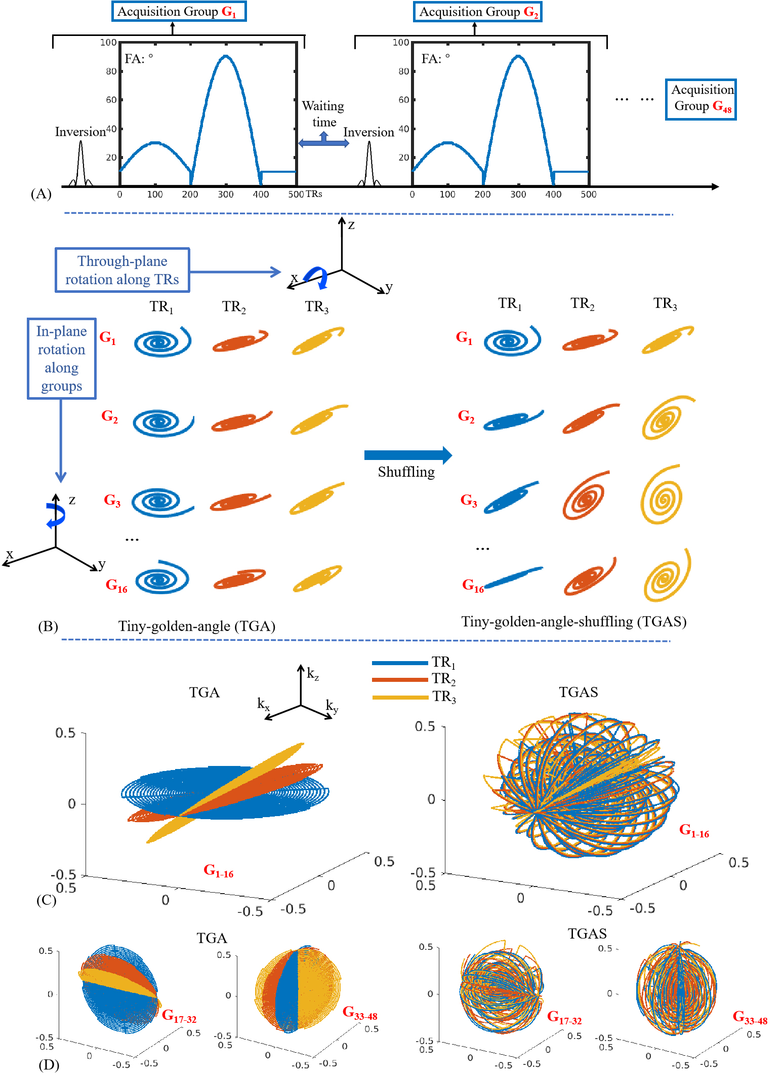
1. (A)
Sequence diagram. (B) Spiral-projection spatiotemporal encoding with left)
the original tiny-golden-angle (TGA) scheme and right) the proposed
tiny-golden-angle-shuffling (TGAS) scheme. (C) and (D) the k-space
coverage of the first 3 TRs for acquisition groups G1-16, G17-32
and G33-48, where through-plane rotation was implemented
around x-, y- and z-axis respectively to achieve multi-axis rotation for better
incoherence.
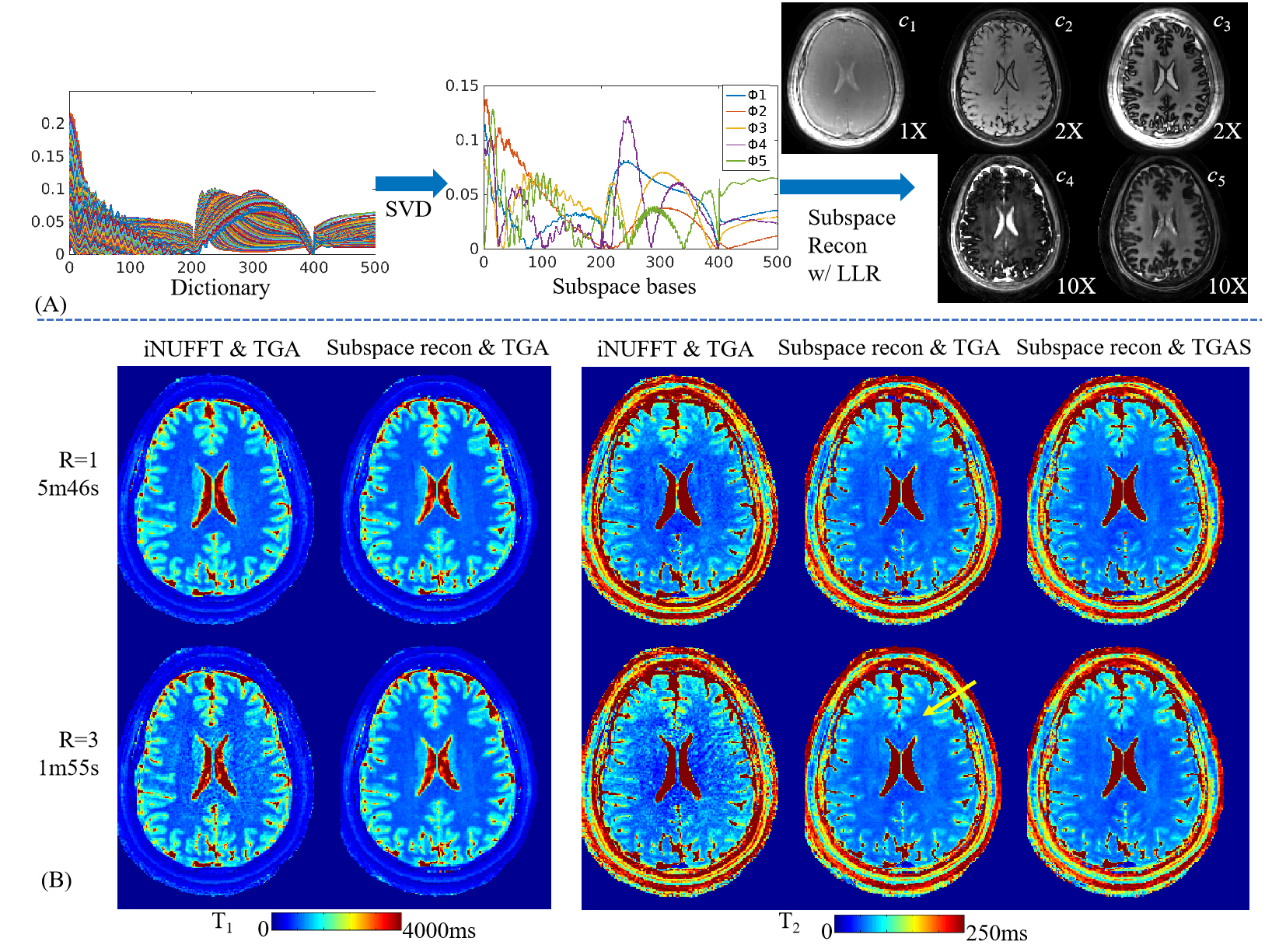
Figure
2. (A) The flow
chart of subspace reconstruction. Five subspace bases were extracted from the MRF
dictionary and used to reconstruct the coefficient maps (at 1×, 2×, 2×, 10×,
and 10× scalings respectively for better visualization). The coefficient maps are
then used to generate the MRF time-series images and dictionary template matching
performed to obtain T1, T2, and PD maps. (B) Comparison
between sliding-window iNUFFT and subspace reconstructions, where T1
& T2 maps from 1-mm isotropic acquisitions at acceleration
factors R=1 & 3 are shown.