Zhehao Hu1,2, Jiayu Xiao1, Xianglun Mao1, Yibin Xie1, Alan Kwan1,3, Xiaoming Bi4, Shlee Song5, Alison Wilcox6, Debiao Li1,2, Anthony Christodoulou1,2, and Zhaoyang Fan1,6,7
1Biomedical Imaging Research Institute, Cedars-Sinai Medical Center, Los Angeles, CA, United States, 2Department of Bioengineering, University of California, Los Angeles, Los Angeles, CA, United States, 3Smidt Heart Institute, Cedars-Sinai Medical Center, Los Angeles, CA, United States, 4Siemens Medical Solutions USA, Inc., Los Angeles, CA, United States, 5Department of Neurology, Cedars-Sinai Medical Center, Los Angeles, CA, United States, 6Department of Radiology, Keck School of Medicine, University of Southern California, Los Angeles, CA, United States, 7Department of Radiation Oncology, Keck School of Medicine, University of Southern California, Los Angeles, CA, United States
1Biomedical Imaging Research Institute, Cedars-Sinai Medical Center, Los Angeles, CA, United States, 2Department of Bioengineering, University of California, Los Angeles, Los Angeles, CA, United States, 3Smidt Heart Institute, Cedars-Sinai Medical Center, Los Angeles, CA, United States, 4Siemens Medical Solutions USA, Inc., Los Angeles, CA, United States, 5Department of Neurology, Cedars-Sinai Medical Center, Los Angeles, CA, United States, 6Department of Radiology, Keck School of Medicine, University of Southern California, Los Angeles, CA, United States, 7Department of Radiation Oncology, Keck School of Medicine, University of Southern California, Los Angeles, CA, United States
Imaging of cardiac anatomy is important in diagnosis and procedural planning. An MR MultiTasking based 3D Multi-dimensional Assessment of Cardiovascular System (MT-MACS) technique is proposed for the assessment of the whole cardiac structures and great thoracic vessels.
Figure 2. Example images generated
by the proposed MT-MACS from a 38-year-old healthy subject. Based on the dual-echo acquisition scheme, MT-MACS can achieve water/fat separation and provide
water-only and fat-only images. For water-only images, three out of 300 image contrasts
are selected for bright-blood, dark-blood and gray-blood imaging, respectively. For
each image contrast, corresponding cardiac phase-resolved cine series can also be
generated to assess the cardiac function. Coronal and transverse views of the fat images, as well as cine fat series, are also displayed.
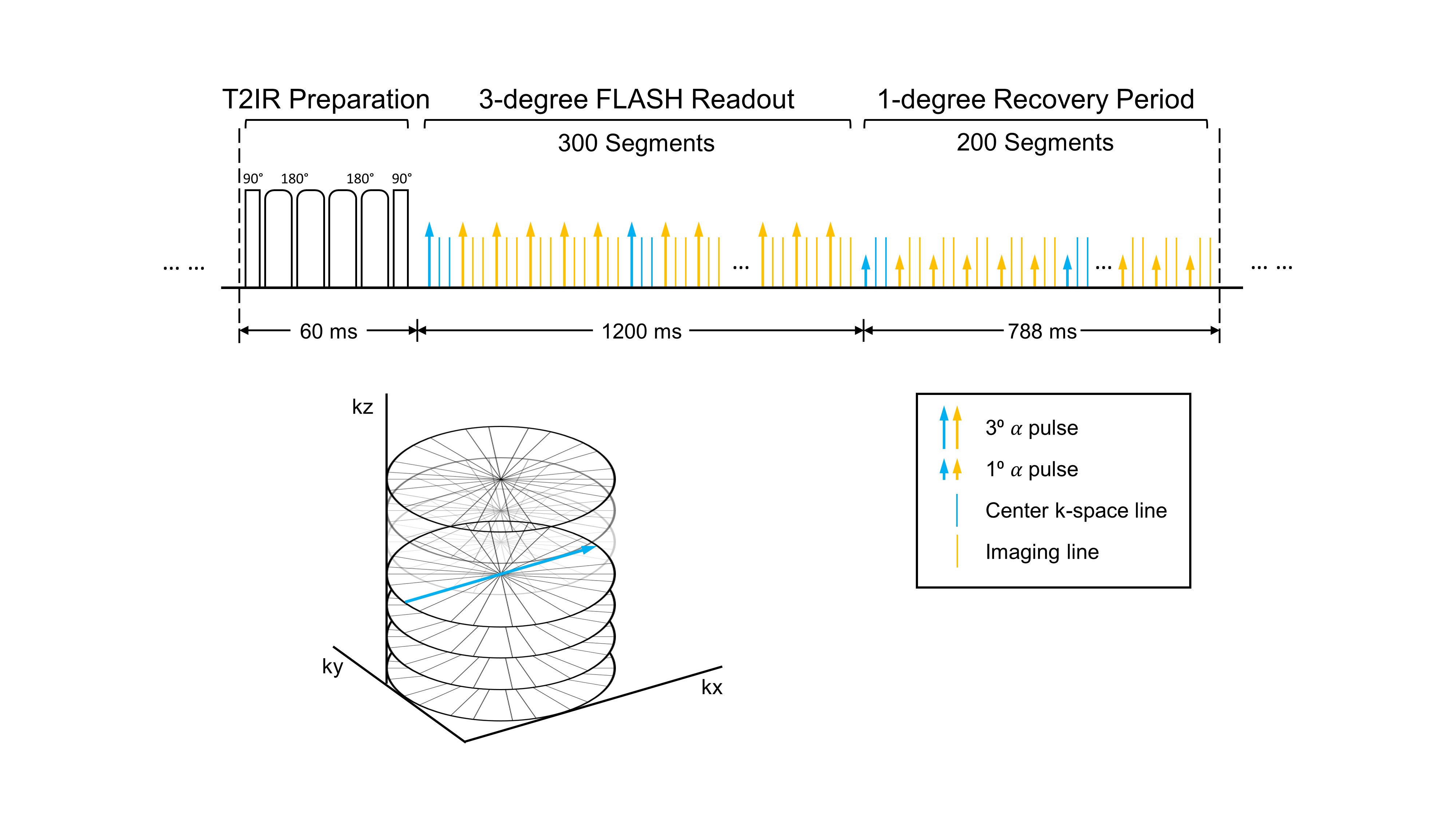
Figure 1. Pulse sequence diagram for
MT-MACS and corresponding k-space sampling pattern for auxiliary data. T2-prepared
inversion recovery (T2IR) magnetization preparations are applied at constant
intervals followed by dual-echo FLASH readouts. RF pulse flip angles following each T2IR are 3⁰ for the
first 300 segments, and 1⁰ for next 200 segments to allow for greater magnetization recovery. Auxiliary data are interleaved with imaging data every
6 readouts and are collected at the 0⁰ radial spoke of the center partition.