Peter Jeon1, Michael MacKinley2, Kara Dempster3, Dickson Wong4, Lena Palaniyappan1,2,5,6, and Jean Theberge1,5,7
1Medical Biophysics, Western University, London, ON, Canada, 2Neuroscience, Western University, London, ON, Canada, 3Psychiatry, Dalhousie University, Halifax, NS, Canada, 4Schulich School of Medicine and Dentistry, Western University, London, ON, Canada, 5Psychiatry, Western University, London, ON, Canada, 6Robarts Research Institute, London, ON, Canada, 7Lawson Health Research Institute, London, ON, Canada
1Medical Biophysics, Western University, London, ON, Canada, 2Neuroscience, Western University, London, ON, Canada, 3Psychiatry, Dalhousie University, Halifax, NS, Canada, 4Schulich School of Medicine and Dentistry, Western University, London, ON, Canada, 5Psychiatry, Western University, London, ON, Canada, 6Robarts Research Institute, London, ON, Canada, 7Lawson Health Research Institute, London, ON, Canada
Functional
MRS at 7T using a color-word Stroop stimulus reveals differences in glutathione
dynamics between first-episode schizophrenia and healthy control groups in the
anterior cingulate cortex.
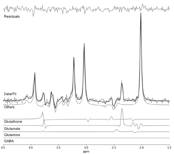
Figure 1: Spectral
fit of one subject during the Rest period (baseline). Metabolites included in
the fitting template were: alanine, aspartate, choline, creatine, GABA,
glucose, glutamate, glutamine, glutathione, glycine, lactate, myo-inositol,
N-acetyl aspartate, N-acetyl aspartyl glutamate, phosphorylethanolamine,
scyllo-inositol, and taurine. Macromolecules were not included due to the long
echo time used.
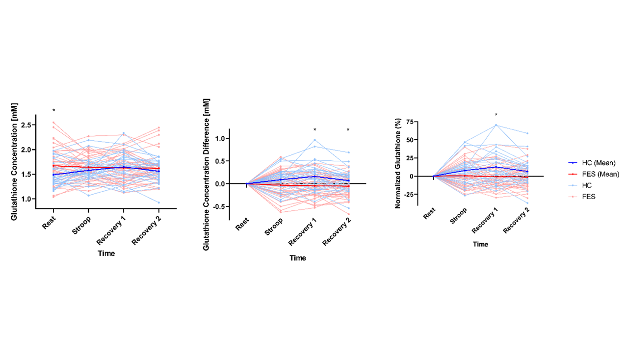
Figure
2: Glutathione (GSH) dynamics. Plots of (a) mean GSH concentration
[mM], (b) mean GSH concentration difference [mM] relative to baseline (‘Rest’),
and (c) mean GSH percentage change (%) normalized to baseline (‘Rest’). Solid
blue, solid red, light blue, and pink lines represent mean HC, mean FES,
individual HC, and individual FES values, respectively. Asterisks above time
points indicate significant difference between group.