Xianjun Li1, Congcong Liu1, Mustafa Salimeen1, Miaomiao Wang1, Mengxuan Li1, Chao Jin1, Xiaocheng Wei1, and Jian Yang1
1Department of Radiology, The First Affiliated Hospital of Xi’an Jiaotong University, Xi'an, China
1Department of Radiology, The First Affiliated Hospital of Xi’an Jiaotong University, Xi'an, China
The ratio between
diffusivities derived from low and high b-value DTI could provide complementary
information for assessing infant cerebrospinal fluid dynamics. There may exist
potential association between Virchow–Robin space volume and cerebrospinal
fluid dynamics.
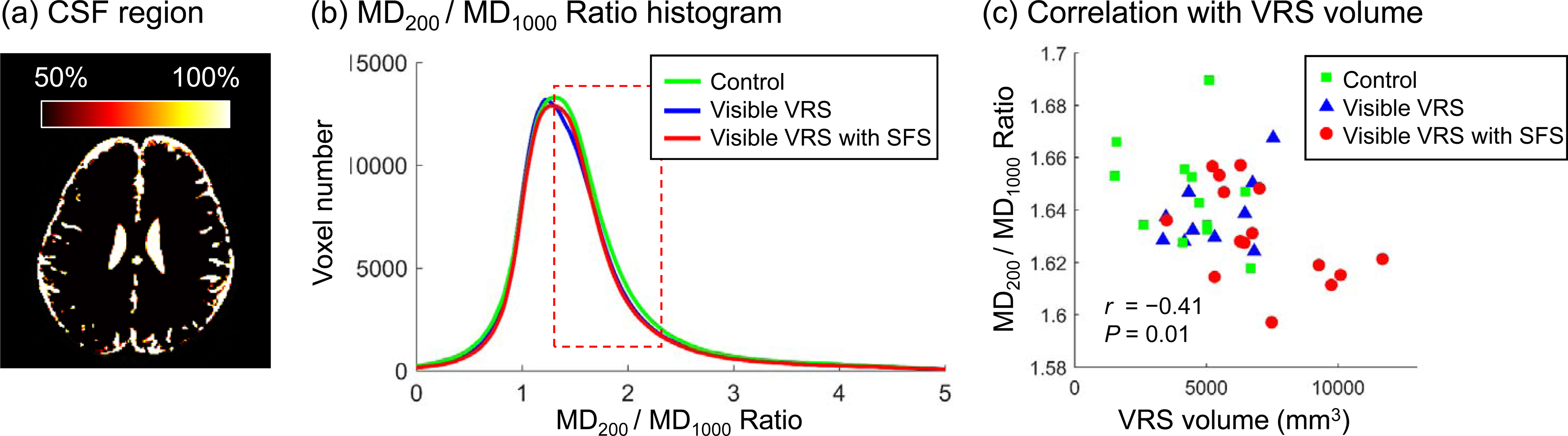
Figure 3. Cerebrospinal fluid (CSF) region (a), MD1000/
MD200 Ratio histogram in the CSF region (b), and correlation between
the VRS volume and the MD1000/ MD200 Ratio (average in ratio
range of 1.3~2.3 in b) (c).
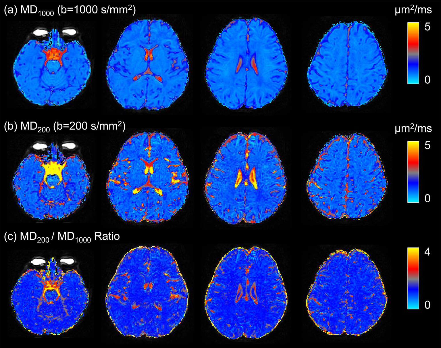
Figure 2. Representative maps of mean diffusivity
based on b value of 1000 s/mm2 (MD1000) (a), mean
diffusivity based on b value of 200 s/mm2 (MD200) (b),
and MD1000/ MD200 Ratio.
