Alexandros Popov1, Raïssa Yebga Hot1, Justine Beaujoin1, Ivy Uszynski1, Fawzi Boumezbeur1, Fabrice Poupon1, Christophe Destrieux2, and Cyril Poupon1
1NeuroSpin (CEA), Paris, France, 2Université de Tours, Tours, France
1NeuroSpin (CEA), Paris, France, 2Université de Tours, Tours, France
In this study, we present the Chenonceau dataset : a novel 11.7T MRI
dataset of the entire human brain, combining an ultra-high resolution
anatomical scan at 100μm with diffusion scans at 200μm using strong
diffusion sensitizations.
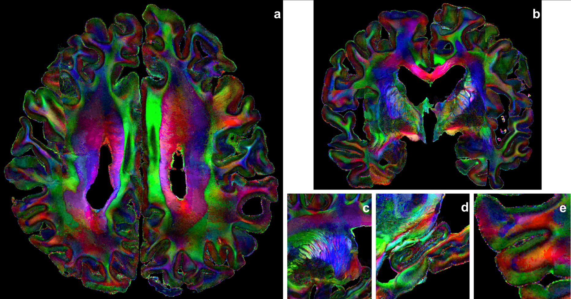
Figure 3: Color-encoded maps computed from the analytical Q-ball model. (a) axial view; (b) coronal view; (c) zoom over the thalamic radiations; (d) zoom over the right hippocampus; (e) zoom over a cortical area.
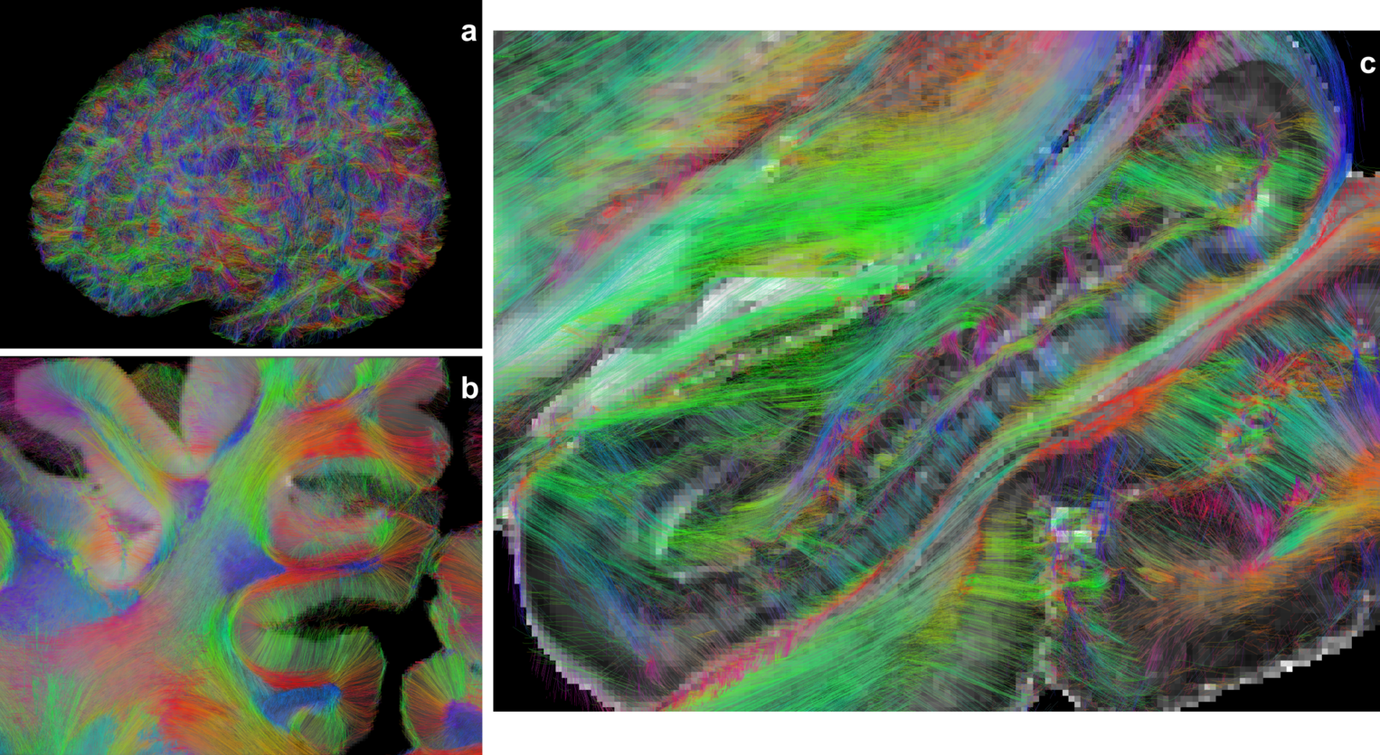
Figure 4: Whole brain connectogram computed from the analytical Q-ball model using a regularized deterministic fiber tracking method; (a) 3d view of the whole connectogram; (b) zoom in frontal lobe superimposed onto the 200μm GFA map, revealing dense and finely structured tracts entering the cortical ribbon; (c) zoom in the right hippocampus superimposed onto the 200μm GFA map, revealing its complex inner fiber organization.