Hecong Qin1,2, Shuyu Tang1, Andrew Riselli1, Robert A. Bok1, Romelyn Delos Santos1, Mark Van Criekinge1, Jeremy W. Gordon1, Rahul Aggarwal3, Evelyn Escobar1, Rui Chen4, Chunxin Tracy Zhang5, Gregory Goddard5, Albert Chen4, Galen Reed4, Ruscitto M. Daniel5, Renuka Sriram1, James Slater1, Peder E.Z. Larson1,2, Daniel B. Vigneron1,2, and John Kurhanewicz1,2
1Radiology and Biomedical Imaging, University of California, San Francisco, San Francisco, CA, United States, 2Graduate Program in Bioengineering, UC Berkeley – UCSF, San Francisco, CA, United States, 3Medicine, University of California, San Francisco, San Francisco, CA, United States, 4GE Healthcare, Waukesha, WI, United States, 5GE Research, Niskayuna, NY, United States
1Radiology and Biomedical Imaging, University of California, San Francisco, San Francisco, CA, United States, 2Graduate Program in Bioengineering, UC Berkeley – UCSF, San Francisco, CA, United States, 3Medicine, University of California, San Francisco, San Francisco, CA, United States, 4GE Healthcare, Waukesha, WI, United States, 5GE Research, Niskayuna, NY, United States
We developed a formulation and standard operating procedure that can reproducibly generate sterile hyperpolarized 13C pyruvate and urea solutions for human injection, as well as a novel imaging approach for simultaneous metabolic and perfusion imaging.

Figure 1. Imaging mechanism: hyperpolarized 13C pyruvate and urea are intravenously injected in a single bolus, then leave the vasculature and enter exocellular space. Pyruvate can enter the cell and be metabolized into lactate, alanine, or CO2/bicarbonate, evaluating multiple metabolic fluxes, whereas urea is predominately extracellular and metabolic inactive, serving as a perfusion contrast agent. (Ktrans, vascular transfer constant; MCT, monocarboxylate transporter; LDH, lactate dehydrogenase; ALT, alanine transaminase; PDH, pyruvate dehydrogenase).
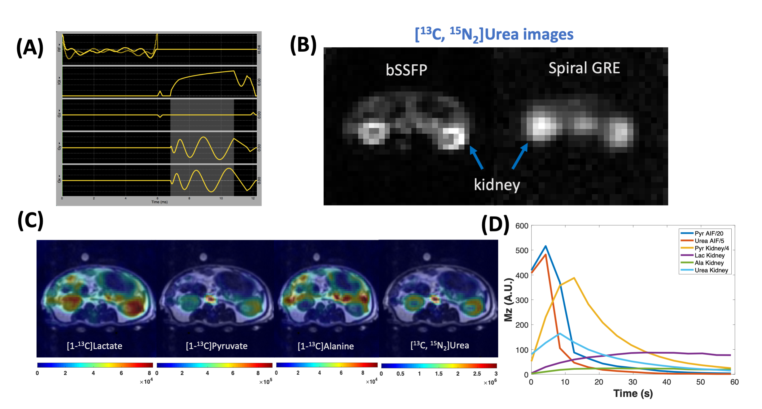
Figure 5. A balanced steady-state free precession (bSSFP) sequence for hyperpolarized (HP) 13C urea (A, highlighted area indicates readout duration) produced superior image qualities (SNR, resolution, sharpness) than a single shot spiral gradient echo (GRE) sequence (B). (C): Representative images of sum HP 13C signal of a healthy rat overlaid on T2 weight images: flip angels of lactate, alanine, pyruvate, and urea are 30°, 30°, 8°, ± 25°; 2.5 × 2.5 × 21mm spatial resolution; 4.2s temporal resolution. (D) HP signal dynamics in the aorta and kidneys of a healthy rat.
